. 2021 Nov 12;13(11):2265.
doi: 10.3390/v13112265.

Rift Valley Fever Virus Propagates in Human Villous Trophoblast Cell Lines and Induces Cytokine mRNA Responses Known to Provoke Miscarriage

Affiliations

Rift Valley Fever Virus Propagates in Human Villous Trophoblast Cell Lines and Induces Cytokine mRNA Responses Known to Provoke Miscarriage

Yong-Dae Gwon et al. Viruses. .

Abstract

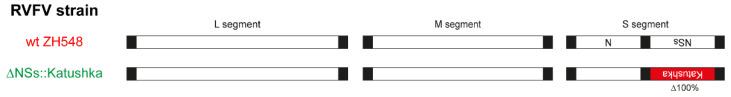
The mosquito-borne Rift Valley fever (RVF) is a prioritised disease that has been listed by the World Health Organization for urgent research and development of counteraction. Rift Valley fever virus (RVFV) can cause a cytopathogenic effect in the infected cell and induce hyperimmune responses that contribute to pathogenesis. In livestock, the consequences of RVFV infection vary from mild symptoms to abortion. In humans, 1-3% of patients with RVFV infection develop severe disease, manifested as, for example, haemorrhagic fever, encephalitis or blindness. RVFV infection has also been associated with miscarriage in humans. During pregnancy, there should be a balance between pro-inflammatory and anti-inflammatory mediators to create a protective environment for the placenta and foetus. Many viruses are capable of penetrating that protective environment and infecting the foetal-maternal unit, possibly via the trophoblasts in the placenta, with potentially severe consequences. Whether it is the viral infection per se, the immune response, or both that contribute to the pathogenesis of miscarriage remains unknown. To investigate how RVFV could contribute to pathogenesis during pregnancy, we infected two human trophoblast cell lines, A3 and Jar, representing normal and transformed human villous trophoblasts, respectively. They were infected with two RVFV variants (wild-type RVFV and RVFV with a deleted NSs protein), and the infection kinetics and 15 different cytokines were analysed. The trophoblast cell lines were infected by both RVFV variants and infection caused upregulation of messenger RNA (mRNA) expression for interferon (IFN) types I-III and inflammatory cytokines, combined with cell line-specific mRNA expression of transforming growth factor (TGF)-β1 and interleukin (IL)-10. When comparing the two RVFV variants, we found that infection with RVFV lacking NSs function caused a hyper-IFN response and inflammatory response, while the wild-type RVFV suppressed the IFN I and inflammatory response. The induction of certain cytokines by RVFV infection could potentially lead to teratogenic effects that disrupt foetal and placental developmental pathways, leading to birth defects and other pregnancy complications, such as miscarriage.

Keywords: cytokine; human villous trophoblast; inflammatory cytokines; interferon; miscarriage; rift valley fever virus.


Conflict of interest statement

The authors declare no conflict of interest. The funders had no role in the design of the study; in the collection, analyses, or interpretation of data; in the writing of the manuscript; or in the decision to publish the results.

Figures

Figure 1
Figure 1
Schematics of the Rift Valley fever virus (RVFV) strains used in this study. The image shows the genomes of two different RVFV strains. The ΔNSs::Katushka strain was derived from the wild-type (wt) ZH548 strain by replacement of the NSs gene with the far-red fluorescent protein Katushka gene.
Figure 2
Figure 2
Rift Valley fever virus (RVFV) Gn protein expression in the A3 and Jar cell lines. The cell lines were infected at a multiplicity of infection of 1 and the Gn protein was detected by using an anti-RVFV Gn monoclonal antibody, and a secondary anti-mouse antibody conjugated to Alexa Fluor 488. Cytation 5 Cell Imaging Multi-Mode Reader identified green fluorescent protein (GFP)- or 4′,6-diamidino-2-phenylindole (DAPI)-expressing cells and quantified the fluorescence intensity in each well. Experiments were performed in triplicate and repeated twice. The bar plus error bar indicates the mean ± standard deviation. Statistical significance was determined by multiple t-test. p values are indicated (NS = not significant).
Figure 3
Figure 3
Interferon (IFN) mRNA response in A3 (immortalised human normal trophoblasts) and Jar (human choriocarcinoma) cell lines infected with two different Rift Valley fever virus (RVFV) strains. The cell lines were infected at a multiplicity of infection of 1 and cell lysates were harvested at 0 h and at 6 or 24 h post-infection (hpi). Total cellular RNA was extracted from harvested cell lysate and used for detection of IFNα1 (A), IFNβ1 (B), IFNγ (C), and IFNλ (D) RNA using reverse transcription-quantitative polymerase chain reaction (RT-qPCR) with TaqMan® FAM/MGB probe assays. Experiments were performed in triplicate and repeated twice. The symbol plus error bar indicates the mean ± standard deviation. Statistical significance was determined by one-way analysis of variance (ANOVA) plus Dunnett’s post hoc analysis. The data from 24 hpi was used for multiple group comparisons and the significance of p values is indicated by the asterisks; red line = mock vs. wt ZH548; green line = mock vs. ΔNSs::Katushka; black line = wt ZH548 vs. ΔNSs::Katushka (* p < 0.05, ** p < 0.01, *** p < 0.001, **** p < 0.0001, NS = not significant).
Figure 4
Figure 4
Th2 cytokine mRNA response in A3 cells (immortalised human normal trophoblast) and Jar (human choriocarcinoma) cell lines infected with two different Rift Valley fever virus (RVFV) strains. Both cell lines were infected at a multiplicity of infection of 1 and cell lysates were harvested at 6 or 24 h post-infection (hpi). The total cellular RNA was extracted from harvested cell lysates and used for detection of interleukin (IL)-4 (A) and IL-5 (B) RNA by using reverse transcription-quantitative polymerase chain reaction (RT-qPCR) with TaqMan® FAM/MGB probe assays. Experiments were performed in triplicate and repeated two times with similar results. The symbol plus error bar indicates the mean ± standard deviation. Statistical significance was determined by one-way analysis of variance (ANOVA) plus Dunnett’s post hoc analysis. The data from 24 hpi was used for multiple group comparison and the significance of p values is indicated by the asterisks; red line = mock vs. wt ZH548; green line = mock vs. ΔNSs::Katushka; black line = wt ZH548 vs. ΔNSs::Katushka (**** p < 0.0001, NS = not significant).
Figure 5
Figure 5
The inflammatory cytokine response in A3 (immortalised human normal trophoblast) and Jar (human choriocarcinoma) cell lines infected with two different Rift Valley fever virus (RVFV) strains. Both cell lines were infected at a multiplicity of infection of 1 and cell lysates were harvested at 6 or 24 h post-infection (hpi). The total cellular RNA was extracted from harvested cell lysates and used for detection of the inflammation-associated cytokine messenger RNA (mRNA) (interleukin (IL)-1β (A), IL-6 (B), IL-8 (C), and tumour necrosis factor (TNF)-α (D)) by using reverse transcription-quantitative polymerase chain reaction (RT-qPCR) with TaqMan® FAM/MGB probe assays. Experiments were performed in triplicate and repeated twice. The symbol plus error bar indicates the mean ± standard deviation. Statistical significance was determined by one-way analysis of variance (ANOVA) plus Dunnett’s post hoc analysis. The data from 24 hpi was used for multiple group comparison the significance of p values is indicated by the asterisks; red line = mock vs. wt ZH548; green line = mock vs. ΔNSs::Katushka; black line = wt ZH548 vs. ΔNSs::Katushka (* p < 0.05, **** p < 0.0001, NS = not significant).
Figure 6
Figure 6
Differential messenger RNA (mRNA) expression of immunosuppressive/T regulatory cell (Treg) cytokines in A3 (immortalised human normal trophoblast) and Jar (human choriocarcinoma) cell lines infected with two different Rift Valley fever virus (RVFV) strains. Both cell lines were infected at a multiplicity of infection of 1 and cell lysates were harvested at 6 or 24 h post-infection (hpi). The total cellular RNA was extracted from harvested cell lysates and used for detection of interleukin (IL)-10 (A) and transforming growth factor (TGF)-β1 (B) mRNA by using reverse transcription-quantitative polymerase chain reaction (RT-qPCR) with TaqMan® FAM/MGB probe assays. Experiments were performed in triplicate and repeated two times with similar results. The symbol plus error bar indicates the mean ± standard deviation. Statistical significance was determined by one-way analysis of variance (ANOVA) plus Dunnett’s post hoc analysis. The data from 24 hpi was used for multiple group comparison and the significance of p values is indicated by the asterisks; red line = mock vs. wt ZH548; green line = mock vs. ΔNSs::Katushka; black line = wt ZH548 vs. ΔNSs::Katushka (*** p < 0.001, **** p < 0.0001, NS = not significant).
Figure 7
Figure 7
Programmed cell death and survival-related gene expression in in A3 (immortalised human normal trophoblast) and Jar (human choriocarcinoma) cell lines infected with two different Rift Valley fever virus (RVFV) strains. Both cell lines were infected at a multiplicity of infection of 1 and cell lysates were harvested at 6 or 24 h post-infection (hpi). The total cellular RNA was extracted from harvested cell lysate and used for detection of programmed cell death and survival-related gene expression of TP53 (A), MAP1LC3A (B), and NK-κB1 (C), by using reverse transcription-quantitative polymerase chain reaction (RT-qPCR) with TaqMan® FAM/MGB probe assays. Experiments were performed in triplicate and repeated two times with similar results. The symbol plus error bar indicates the mean ± standard deviation. Statistical significance was determined by one-way analysis of variance (ANOVA) plus Dunnett’s post hoc analysis. The data from 24 hpi was used for multiple group comparison and the significance of p values is indicated by the asterisks; red line = mock vs. wt ZH548; green line = mock vs. ΔNSs::Katushka; black line = wt ZH548 vs. ΔNSs::Katushka (* p < 0.05, ** p < 0.01, *** p < 0.001, **** p < 0.0001, NS = not significant).

References

    1. World Health Organization. [(accessed on 14 August 2020)]. Available online: https://www.who.int/activities/prioritizing-diseases-for-research-and-de....
    1. Bouloy M., Weber F. Molecular biology of rift valley Fever virus. Open Virol. J. 2010;4:8–14. doi: 10.2174/1874357901004010008. - DOI - PMC - PubMed
    1. Bird B.H., Ksiazek T.G., Nichol S.T., Maclachlan N.J. Rift Valley fever virus. J. Am. Vet. Med. Assoc. 2009;234:883–893. doi: 10.2460/javma.234.7.883. - DOI - PubMed
    1. McMillen C.M., Hartman A.L. Rift Valley fever in animals and humans: Current perspectives. Antivir. Res. 2018;156:29–37. doi: 10.1016/j.antiviral.2018.05.009. - DOI - PMC - PubMed
    1. Mansfield K.L., Banyard A.C., McElhinney L., Johnson N., Horton D.L., Hernandez-Triana L.M., Fooks A.R. Rift Valley fever virus: A review of diagnosis and vaccination, and implications for emergence in Europe. Vaccine. 2015;33:5520–5531. doi: 10.1016/j.vaccine.2015.08.020. - DOI - PubMed

Publication types

MeSH terms