. 2021 Nov 17;13(11):2297.
doi: 10.3390/v13112297.

Natural History of Aerosol-Induced Ebola Virus Disease in Rhesus Macaques

Affiliations

Natural History of Aerosol-Induced Ebola Virus Disease in Rhesus Macaques

Isaac Downs et al. Viruses. .

Abstract

Ebola virus disease (EVD) is a serious global health concern because case fatality rates are approximately 50% due to recent widespread outbreaks in Africa. Well-defined nonhuman primate (NHP) models for different routes of Ebola virus exposure are needed to test the efficacy of candidate countermeasures. In this natural history study, four rhesus macaques were challenged via aerosol with a target titer of 1000 plaque-forming units per milliliter of Ebola virus. The course of disease was split into the following stages for descriptive purposes: subclinical, clinical, and decompensated. During the subclinical stage, high levels of venous partial pressure of carbon dioxide led to respiratory acidemia in three of four of the NHPs, and all developed lymphopenia. During the clinical stage, all animals had fever, viremia, and respiratory alkalosis. The decompensatory stage involved coagulopathy, cytokine storm, and liver and renal injury. These events were followed by hypotension, elevated lactate, metabolic acidemia, shock and mortality similar to historic intramuscular challenge studies. Viral loads in the lungs of aerosol-exposed animals were not distinctly different compared to previous intramuscularly challenged studies. Differences in the aerosol model, compared to intramuscular model, include an extended subclinical stage, shortened clinical stage, and general decompensated stage. Therefore, the shortened timeframe for clinical detection of the aerosol-induced disease can impair timely therapeutic administration. In summary, this nonhuman primate model of aerosol-induced EVD characterizes early disease markers and additional details to enable countermeasure development.

Keywords: Ebola; Kikwit; Macaca mulatta; Zaire; aerosol; cytokine storm; natural history; respiratory alkalosis; rhesus macaque; telemetry; viral hemorrhagic fever; virus.


Conflict of interest statement

The authors declare no conflict of interest.

Figures

Figure 1
Figure 1
Viral detection in sera and tissues of EBOV-challenged rhesus macaques. (a) Plaque titer (PFU/mL) of serum for each NHP at specific time points. DPE 0 is marked by a black vertical dot line. The black horizontal dashed line is the average of all animals for DPE 0. (b) EBOV RNA equivalent levels at terminal endpoint of NHPs for respective tissues; lymph node (LN). RNA was amplified using qRT-PCR to identify Ebola virus glycoprotein gene in tissue of each NHP. NHP color designation is the same as (a).
Figure 2
Figure 2
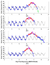
Telemetric temperature detection of aerosol EBOV-challenged rhesus macaques: (a) NHP1, (b) NHP2, (c) NHP3, and (d) NHP4. Vertical grey dotted line marks DPE 0—BL (baseline), ♦ marks fever (>1.5 °C over baseline) for longer than 2 h, ♦ marks LS (values significantly lower: <3.0 SD from corresponding baseline) for longer than 2 h, ♦ marks temperature (°C) for longer than 2 h, and ♦ marks hyperpyrexia (>3.0 °C over baseline) for longer than 2 h.
Figure 3
Figure 3
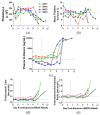
Time course of blood gases and constituents of EBOV-challenged rhesus macaques. Detection of (a) partial pressure of carbon dioxide (pCO2), (b) total CO2, (c) bicarbonate, (d) base excess of extracellular fluid, (e) pH, and (f) lactate were determined in whole blood using i-Stat CG4+. DPE 0 is marked by a grey vertical dot line. The horizontal dashed line is the average of all animals for DPE 0.
Figure 4
Figure 4
Time course of serum liver enzyme activity of EBOV-challenged rhesus macaques. Serum levels of (a) alanine aminotransferase, (b) aspartate aminotransferase, (c) gamma-glutamyl transferase, and (d) alkaline phosphatase were detected using Piccolo chemistry. DPE 0 is marked by a black vertical dot line. The black horizontal dashed line is the average of all animals for DPE 0.
Figure 5
Figure 5
Time course analysis of red blood cell mass and white blood cells from aerosol EBOV-challenged cynomolgus rhesus macaques: (a) hematocrit percentage, (b) number of red blood cells, (c) hemoglobin concentration, (d) number of white blood cells, (e) number of lymphocytes, (f) monocytes, (g) number of neutrophils, (h) number of basophils, and (i) number of eosinophils detected per microliter of blood. DPE 0 is marked by a black vertical dot line. Black horizontal dashed line is average of all animals for DPE 0.
Figure 6
Figure 6
Analysis of immune responses in blood from aerosol EBOV-challenged rhesus macaques. Cytokine analyses of blood—(a) IFNγ, (b) IL-18, and (c) MCP-1—were performed on samples collected 3 days prior to exposure and DPE 0–10. DPE 0 is marked by a black vertical dot line. Black horizontal dashed line is the average of all animals for DPE 0.
Figure 7
Figure 7
Assessment of platelets and clot formation in the blood of aerosol EBOV-challenged rhesus macaques. Blood (a) platelets, (b) mean platelet volume, (c) plasma D-dimers, (d) prothrombin time, and (e) international normalized ratio were analyzed in samples collected 3 days prior to exposure and DPE 0–10. DPE 0 is marked by a black vertical dot line. The black horizontal dashed line is the average of all animals for DPE 0.
Figure 8
Figure 8
Natural history of aerosol-induced EVD in rhesus macaques. The subclinical stage of disease progression is defined by absence of fever between DPE 0 and 4. The clinical stage of disease progression is defined by fever, as observed on DPE 5 in the current study and the serial sacrifice study by Twenhafel et al. 2012. Multiple tissues (qRT-PCR EBOV-positive) are in descending order: inguinal LN, liver, spleen, tracheobronchial LN, lung, axillary LN, mandibular LN, kidney, adrenal gland, mesenteric LN, and bone marrow. The decompensated stage of the natural history study was identified by coagulopathy, cytokine storm, liver injury, kidney malfunction, and mean clinical score ≥ 2.0. Macular rashes were found on the face, pinna, arms, and legs of animals. Here, the aerosol-induced EVD had an extended subclinical stage, shortened clinical stage, and general decompensated stage when compared to historic intramuscular studies noted in Figure S15 (lymph node = LN; solid black line = subclinical stage; solid gray arrow = start of clinical stage; solid gray line = clinical stage; and dashed gray arrow = start of decompensated stage). * Asterisk pertains to Twenhafel et al. 2012.

References

    1. Bibby K., Fischer R.J., Casson L.W., Stachler E., Haas C.N., Munster V.J. Persistence of Ebola Virus in Sterilized Wastewater. Environ. Sci. Technol. Lett. 2015;2:245–249. doi: 10.1021/acs.estlett.5b00193. - DOI - PMC - PubMed
    1. Gordon T.B., Hayward J.A., Marsh G.A., Baker M.L., Tachedjian G. Host and Viral Proteins Modulating Ebola and Marburg Virus Egress. Viruses. 2019;11:25. doi: 10.3390/v11010025. - DOI - PMC - PubMed
    1. Noda T., Sagara H., Suzuki E., Takada A., Kida H., Kawaoka Y. Ebola virus VP40 drives the formation of virus-like filamentous particles along with GP. J. Virol. 2002;76:4855–4865. doi: 10.1128/JVI.76.10.4855-4865.2002. - DOI - PMC - PubMed
    1. Johnson E., Jaax N., White J., Jahrling P. Lethal experimental infections of rhesus monkeys by aerosolized Ebola virus. Int. J. Exp. Pathol. 1995;76:227–236. - PMC - PubMed
    1. WHO Ebola Virus Disease. [(accessed on 13 September 2020)]. Available online: https://www.who.int/news-room/fact-sheets/detail/ebola-virus-disease.

Publication types