. 2021 Nov 18;13(11):2299.
doi: 10.3390/v13112299.

The Roles of Amphibian (Xenopus laevis) Macrophages during Chronic Frog Virus 3 Infections

Affiliations

The Roles of Amphibian (Xenopus laevis) Macrophages during Chronic Frog Virus 3 Infections

Muhammad Riadul Haque Hossainey et al. Viruses. .

Abstract

Infections by Frog Virus 3 (FV3) and other ranavirus genus members are significantly contributing to global amphibian decline. The Xenopus laevis frog is an ideal research platform upon which to study the roles of distinct frog leukocyte populations during FV3 infections. Frog macrophages (MΦs) are integrally involved during FV3 infection, as they facilitate viral dissemination and persistence but also participate in immune defense against this pathogen. In turn, MΦ differentiation and functionality depend on the colony-stimulating factor-1 receptor (CSF-1R), which is ligated by CSF-1 and iterleukin-34 (IL-34) cytokines. Our past work indicated that X. laevis CSF-1 and IL-34 give rise to morphologically and functionally distinct frog MΦ subsets, and that these CSF-1- and IL-34-MΦs respectively confer susceptibility and antiviral resistance to FV3. Because FV3 targets the frog kidneys and establishes chronic infections therein, presently we examined the roles of the frog CSF-1- and IL-34-MΦs in seeding and maintaining these chronic kidney infections. Our findings indicate that the frog CSF-1-MΦs result in more prominent kidney FV3 infections, which develop into greater reservoirs of lingering FV3 marked by infiltrating leukocytes, fibrosis, and overall immunosuppressive states. Moreover, the antiviral effects of IL-34-MΦs are short-lived and are lost as FV3 infections progress.

Keywords: amphibian; interferons; intestine; myeloid cells; ranavirus.


Conflict of interest statement

The authors declare no conflict of interest.

Figures

Figure 1
Figure 1
CSF-1-MΦ-administered frogs succumb to FV3 infections. X. laevis were injected ip with 2.5 μg of rCSF-1 or rIL-34 in APBS or equal volumes of a recombinant control (r-ctrl) and three days later infected ip with FV3 (5 × 10⁵ PFU). Animal survival was monitored over the course of 50 days, n = 15 per treatment group.
Figure 2
Figure 2
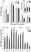
FV3 loads and gene expression analyses in control, CSF-1- and IL-34-MΦ-enriched frogs. X. laevis were injected ip with 2.5 μg of rCSF-1 or rIL-34 in APBS or equal volumes of empty vector control (r-ctrl) and three days later infected ip with FV3 (5 × 10⁵ PFU). At designated times, animals were sacrificed, and their kidneys were examined by qPCR for (A) FV3 DNA loads (n = 6), (B) by plaque assays for FV3 infectious viral particle content per kidney (n = 5) at 7 and 21 dpi, and (C) by qPCR for FV3 gene expression of icp18, rad2 and mcp genes (n = 6). The results are means ± SE. The letters above head bars indicate statistical groups, with each letter representing those treatment groups that are not statically different from each other and distinct letters indicating treatment groups that are significantly different. Asterisks above lines (*) denote statistical differences between the treatment groups denoted by the lines, p < 0.05.
Figure 3
Figure 3
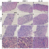
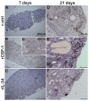
CSF-1-MΦ-enriched, FV3 infected frogs possess greater kidney tissue damage and infiltrating leukocytes. X. laevis were injected ip with 2.5 μg of rCSF-1 (B,E,H,K) or rIL-34 (C,F,I,L) in APBS or equal volumes of a recombinant control (r-ctrl; (A,D,G,J)) and three days later infected ip with FV3 (5 × 10⁵ PFU). At designated times, animals were sacrificed, and their kidneys were processed for histology and stained with hematoxylin & eosin and examined by microscopy. Fibrosis (f) and leukocytes (arrows) are indicated, and the images are representative of sections from four mock-infected and five infected animals per treatment group (n = 4 for uninfected controls and n = 5 for FV3-infected groups) individual animal kidneys from respective treatment groups and times.
Figure 4
Figure 4
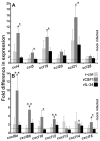
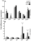
Kidneys from CSF-1-MΦ-enriched, and chronically FV3-infected frogs possess greater expression of chemokine genes. X. laevis were injected ip with 2.5 μg of rCSF-1 or rIL-34 in APBS or equal volumes of empty vector control (r-ctrl) and three days later infected ip with FV3 (5 × 10⁵ PFU) or mock-infected with ip APBS injections. After 28 days of infection, animals were sacrificed, and their kidneys were examined by qPCR for expression of (A) CC and (B) CXC motif chemokine genes. The results are means ± SE (n = 6). All expression was normalized against mock-infected controls, with average baseline (uninfected) expression indicated by horizontal lines. Asterisks (*) indicate significant increase in gene expression above mock-infected controls and asterisks above lines (*) denote statistical differences between the treatment groups denoted by the lines, p < 0.05.
Figure 5
Figure 5
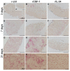
The kidneys of CSF-1-MΦ-enriched, FV3 infected frogs possess greater infiltration of granulocytes. X. laevis were injected ip with 2.5 μg of rCSF-1 (B,E,H,K) or rIL-34 (C,F,I,L) in APBS or equal volumes of a recombinant vector control (r-ctrl; (A,D,G,J); inset a panel: uninfected) and three days later infected ip with FV3 (5 × 10⁵ PFU). At designated times, animals were sacrificed, and their kidneys were processed for histology and examined by NASDCl-specific esterase (Leder) stain, with granulocytes staining pink. The images are representative of sections from kidneys of four mock-infected and five infected animals per treatment group (n = 4 for uninfected controls and n = 5 for FV3-infected groups). Inset panel in (A), denoted (a) is representative of kidneys from mock-infected animals.
Figure 6
Figure 6
The kidneys of CSF-1-MΦ-enriched, FV3 infected frogs possess greater infiltration of MΦs. X. laevis were injected ip with 2.5 μg of rCSF-1 (B, inset b, E) or rIL-34 (C,F) in APBS or equal volumes of a recombinant control (r-ctrl; A,D) and three days later infected ip with FV3 (5 × 10⁵ PFU). At designated times, animals were sacrificed, and their kidneys were processed for histology and examined by α-Naphthyl Acetate (non-specific esterase; Sigma-Aldrich) stain, with MΦ-lineage cells staining brownish-black. The images are representative of sections from kidneys of five infected animals per treatment group (n = 5 for FV3-infected groups). Inset panel in (B), denoted as (b) is higher magnification of part of the same section.
Figure 7
Figure 7
Kidneys from CSF-1-MΦ-enriched, and chronically FV3-infected frogs possess greater expression of myeloid cell markers and immunosuppressive genes. X. laevis were injected ip with 2.5 μg of rCSF-1 or rIL-34 in APBS or equal volumes of empty vector control (r-ctrl) and three days later infected ip with FV3 (5 × 10⁵ PFU) or mock-infected with ip APBS injections. After 28 days of infection, animals were sacrificed, and their kidneys were examined by qPCR for gene expression of (A) leukocyte markers and (B) cytokines. The results are means ± SE (n = 6). All expression was normalized against mock-infected controls, with average baseline (uninfected) expression indicated by horizontal lines. Asterisks (*) indicate significant increase in gene expression above mock-infected controls and asterisks above lines (*) denote statistical differences between the treatment groups denoted by the lines, p < 0.05.

Similar articles

Cited by

References

    1. Cunningham A.A., Hyatt A.D., Russell P., Bennett P.M. Emerging epidemic diseases of frogs in Britain are dependent on the source of ranavirus agent and the route of exposure. Epidemiol. Infect. 2007;135:1200–1212. doi: 10.1017/S0950268806007679. - DOI - PMC - PubMed
    1. Greer A.L., Berrill M., Wilson P.J. Five amphibian mortality events associated with ranavirus infection in south central Ontario, Canada. Dis. Aquat. Organ. 2005;67:9–14. doi: 10.3354/dao067009. - DOI - PubMed
    1. Zupanovic Z., Lopez G., Hyatt A.D., Green B., Bartran G., Parkes H., Whittington R.J., Speare R. Giant toads Bufo marinus in Australia and Venezuela have antibodies against ‘ranaviruses’. Dis. Aquat. Organ. 1998;32:1–8. doi: 10.3354/dao032001. - DOI - PubMed
    1. Green D.E., Converse K.A., Schrader A.K. Epizootiology of sixty-four amphibian morbidity and mortality events in the USA, 1996–2001. Ann. N. Y. Acad. Sci. 2002;969:323–339. doi: 10.1111/j.1749-6632.2002.tb04400.x. - DOI - PubMed
    1. Duffus A.L., Pauli B.D., Wozney K., Brunetti C.R., Berrill M. Frog virus 3-like infections in aquatic amphibian communities. J. Wildl. Dis. 2008;44:109–120. doi: 10.7589/0090-3558-44.1.109. - DOI - PubMed

Publication types