. 2021 Nov 19;13(11):2308.
doi: 10.3390/v13112308.

Huntingtin-Interacting Protein 1 Promotes Vpr-Induced G2 Arrest and HIV-1 Infection in Macrophages

Affiliations

Huntingtin-Interacting Protein 1 Promotes Vpr-Induced G2 Arrest and HIV-1 Infection in Macrophages

Tomoyuki Murakami et al. Viruses. .

Abstract

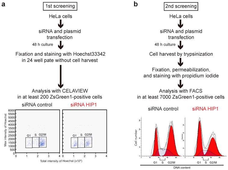
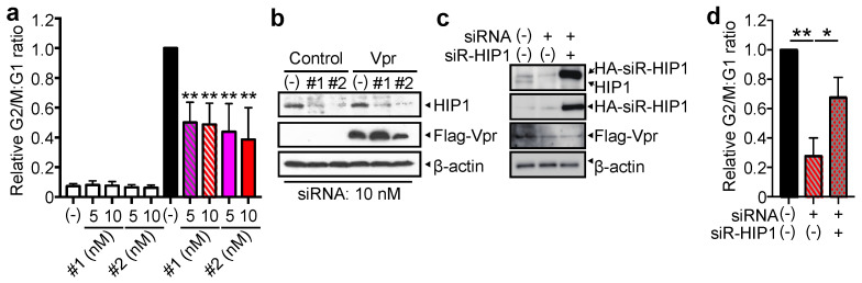
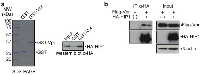
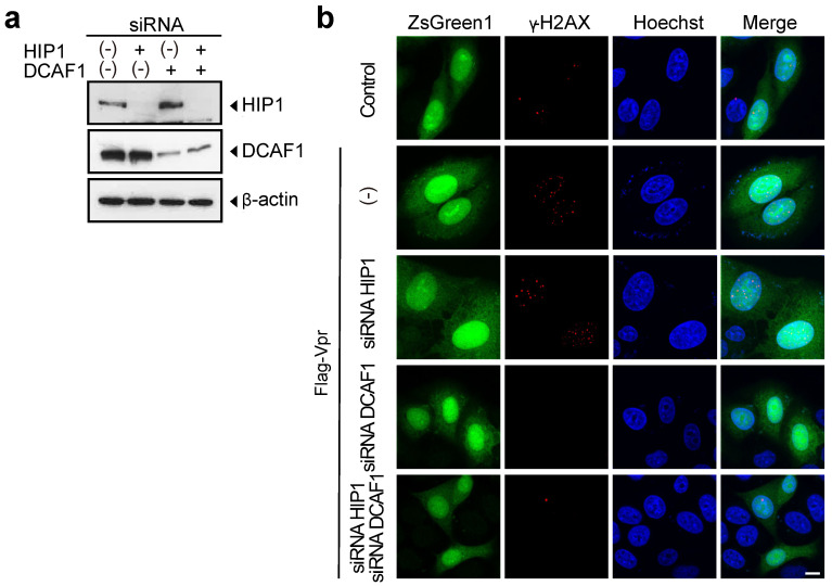
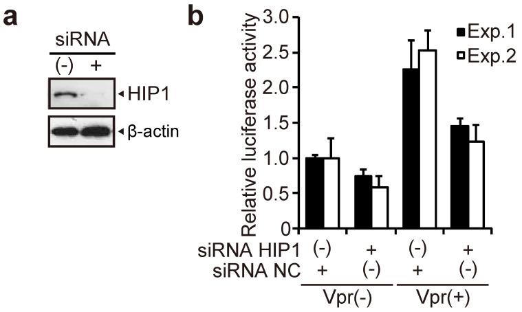
Human immunodeficiency virus type 1 (HIV-1) modulates the host cell cycle. The HIV-1 accessory protein Vpr arrests the cell cycle at the G2 phase in dividing cells, and the ability of Vpr to induce G2 arrest is well conserved among primate lentiviruses. Additionally, Vpr-mediated G2 arrest likely correlates with enhanced HIV-1 infection in monocyte-derived macrophages. Here, we screened small-interfering RNA to reveal candidates that suppress Vpr-induced G2 arrest and identified Huntingtin-interacting protein 1 (HIP1) required for efficient G2 arrest. Interestingly, HIP1 was not essential for Vpr-induced DNA double-strand breaks, which are required for activation of the DNA-damage checkpoint and G2 arrest. Furthermore, HIP1 knockdown suppressed HIV-1 infection in monocyte-derived macrophages. This study identifies HIP1 as a factor promoting Vpr-induced G2 arrest and HIV-1 infection in macrophages.

Keywords: G2 arrest; HIP1; HIV-1; Vpr; macrophage.


Conflict of interest statement

The authors declare no conflict of interest.

Figures

Figure 1
Figure 1
Summary of siRNA screening for candidate suppressors of Vpr-induced G2 arrest. (a) For the initial screening, HeLa cells were co-transfected with pME/F-Vpr-IRES-ZsGreen1 and 100 nM individual siRNAs from the siRNA mini-library or control siRNA. At 48-h post-transfection, cells were fixed and stained with 5 μg/mL Hoechst33342 to measure DNA content in at least 200 ZsGreen1+ cells using CELAVIEW RS100. (b) For secondary screening, HeLa cells were co-transfected with pME/F-Vpr-IRES-ZsGreen1 and control siRNA or 100 nM of individual siRNAs that inhibited Vpr-induced G2 arrest according to the initial screen. At 48-h post-transfection, cells were harvested by trypsinization, fixed, permeabilized, treated with RNase A, and stained with 50 μg/mL propidium iodide to measure DNA content in at least 7000 ZsGreen1+ cells by flow cytometry. The traces on the cell cycle histograms are written by the ModFit LT software automatically.
Figure 2
Figure 2
HIP1 enhances Vpr-induced G2 arrest. (a,b) HeLa cells were co-transfected with pME/F-Vpr-IRES-ZsGreen1 or control pME/F-IRES-ZsGreen1 and 5 or 10 nM either HIP1 (siRNA#1 and #2) or control siRNA and cultured for 48 h. (a) Cells were fixed and stained with 5 μg/mL Hoechst33342 to measure DNA content in ZsGreen1+ cells using CELAVIEW RS100. The relative G2/M:G1 ratio was plotted, and data represent the mean ± SD of three independent experiments. (b) Cells were lysed and subjected to 6% and 15% SDS-PAGE and Western blot using an anti-HIP1 mAb, anti-Flag M2 mAb, and anti-β-actin mAb. (c,d) HeLa cells were co-transfected with pME/F-Vpr-IRES-ZsGreen1 and pCAGGS/HA-siR-HIP1 (carrying synonymous nucleotide mutations at the third codon of the siRNA-targeting site) or control pCAGGS/HA and either 10 nM HIP1 (siRNA#2) or control siRNA and then cultured for 48 h. (c) Cells were lysed and subjected to 6% and 15% SDS-PAGE and Western blot using an anti-HIP1 mAb, anti-Flag M2 mAb, anti-HA mAb, and anti-β-actin mAb. (d) Cells were fixed, permeabilized, stained with the anti-HA mAb, and then with Alexa Fluor 594-conjugated secondary Ab and Hoechst33342. The DNA content of ZsGreen1+ and Alexa Fluor 594+ cells was analyzed by CELAVIEW RS100. The relative G2/M:G1 ratio was plotted, and data represent the mean ± SD of three independent experiments. * p < 0.05, ** p < 0.01 via two-tailed Student’s t-test.
Figure 3
Figure 3
Vpr interacts with HIP1. (a) GST and GST-Vpr were resolved by 12% SDS-PAGE and stained with Coomassie Brilliant Blue (CBB) (left). Glutathione-Sepharose beads combined with the GST-Vpr or GST alone were incubated with purified HA-HIP1 protein. The bound fractions and 10% of the input were analyzed by Western blot using the anti-HA mAb (right). The positions of HA-HIP1, GST-Vpr, and GST are indicated. (b) 293T cells were co-transfected with pME/FVpr and pCAGGS/HA-HIP1 or control pCAGGS/HA. At 48-h post-transfection, cells were lysed, and the lysates were subjected to immunoprecipitation assays using anti-HA agarose and the HA peptide. The bound fractions and inputs were analyzed by Western blotting using the anti-HA polyclonal Ab, anti-Flag polyclonal Ab, and anti-β-actin mAb.
Figure 4
Figure 4
Vpr induces DNA double-strand breaks in HIP1-knockdown cells. (a) HeLa cells were transfected with 5 nM HIP1 (siRNA#2), DCAF1, or a combination of 5 nM HIP1 (siRNA#2) and 5 nM DCAF1 siRNAs. The total amount of siRNA was adjusted to 10 nM with control siRNA. At 48-h post-transfection, cells were lysed and subjected to SDS-PAGE and Western blot using the anti-HIP1 mAb, anti-DCAF1 mAb, and anti-β-actin mAb. The positions of HIP1, DCAF1, and β-actin are indicated. (b) HeLa cells were co-transfected with pME/F-Vpr-IRES-ZsGreen1 or control pME/F-IRES-ZsGreen1 and 5 nM HIP1 (siRNA#2), DCAF1, or a combination of 5 nM HIP1 (siRNA#2) and/or 5 nM DCAF1 siRNAs. The total amount of siRNA was adjusted to 10 nM with control siRNA. At 48-h post-transfection, cells were fixed, permeabilized, and stained with the anti-γ-H2AX mAb, followed by Alexa Fluor 594-conjugated secondary antibody and 5 μg/mL Hoechst33342. Cells were analyzed by a confocal microscopy. Cells showing green fluorescence (ZsGreen1+) and red foci indicated the presence of DNA double-strand breaks, and blue fluorescence indicated nuclei. The scale bar represents 10 μm.
Figure 5
Figure 5
HIP1 enhances HIV-1 infection in macrophages in a Vpr-dependent manner. Monocytes isolated from PBMCs of human healthy donor were subsequently differentiated into macrophages by addition of M-CSF, followed by their transfection with 50 nM HIP1 (siRNA#2; siRNA HIP1) and control siRNA (siRNA NC). (a) At 48-h post-transfection, cells were lysed and subjected to 6% SDS-PAGE and Western blotting using the anti-HIP1 mAb and anti-β-actin mAb. The positions of HIP1 and β-actin are indicated. (b) At 24-h post-transfection, cells were infected with 4 ng p24 VSV-G-pseudotyped-HIV-1 or -HIV-1 ΔVpr. At 6-days post-infection, cells were lysed, and infectivity was determined by measuring luciferase activity. Data represent the mean ± SD of triplicate wells.

Similar articles

Cited by

References

    1. Hashizume C., Kuramitsu M., Zhang X., Kurosawa T., Kamata M., Aida Y. Human immunodeficiency virus type 1 Vpr interacts with spliceosomal protein SAP145 to mediate cellular pre-mRNA splicing inhibition. Microbes Infect. 2007;9:490–497. doi: 10.1016/j.micinf.2007.01.013. - DOI - PubMed
    1. Kuramitsu M., Hashizume C., Yamamoto N., Azuma A., Kamata M., Yamamoto N., Tanaka Y., Aida Y. A novel role for Vpr of human immunodeficiency virus type 1 as a regulator of the splicing of cellular pre-mRNA. Microbes Infect. 2005;7:1150–1160. doi: 10.1016/j.micinf.2005.03.022. - DOI - PubMed
    1. Zhang X., Aida Y. HIV-1 Vpr: A novel role in regulating RNA splicing. Curr. HIV Res. 2009;7:163–168. doi: 10.2174/157016209787581517. - DOI - PubMed
    1. Chutiwitoonchai N., Siarot L., Takeda E., Shioda T., Ueda M., Aida Y. HIV-1 Vpr Abrogates the Effect of TSG101 Overexpression to Support Virus Release. PLoS ONE. 2016;11:e0163100. doi: 10.1371/journal.pone.0163100. - DOI - PMC - PubMed
    1. Kamata M., Nitahara-Kasahara Y., Miyamoto Y., Yoneda Y., Aida Y. Importin-α Promotes Passage through the Nuclear Pore Complex of Human Immunodeficiency Virus Type 1 Vpr. J. Virol. 2005;79:3557–3564. doi: 10.1128/JVI.79.6.3557-3564.2005. - DOI - PMC - PubMed

Publication types

Substances