. 2021 Oct 27;9(11):2234.
doi: 10.3390/microorganisms9112234.

Novel Cytoskeleton-Associated Proteins in Trypanosoma brucei Are Essential for Cell Morphogenesis and Cytokinesis

Affiliations

Novel Cytoskeleton-Associated Proteins in Trypanosoma brucei Are Essential for Cell Morphogenesis and Cytokinesis

Marina Schock et al. Microorganisms. .

Abstract

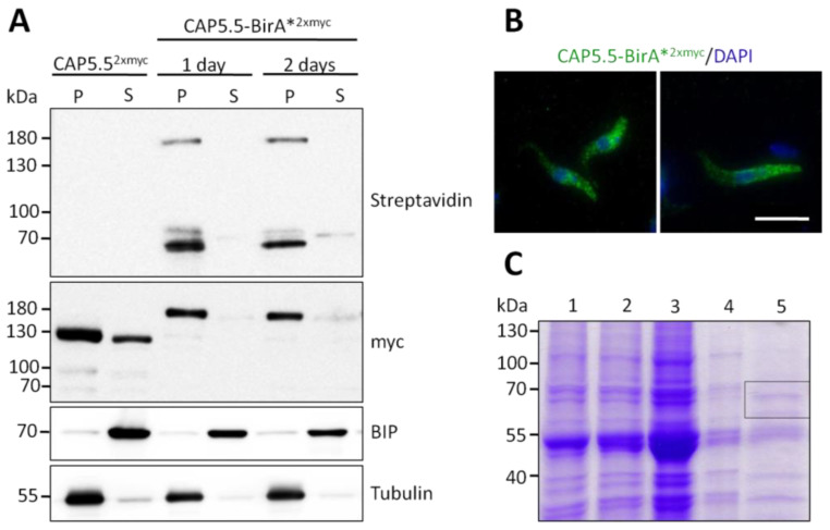
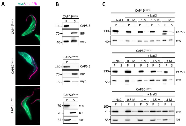
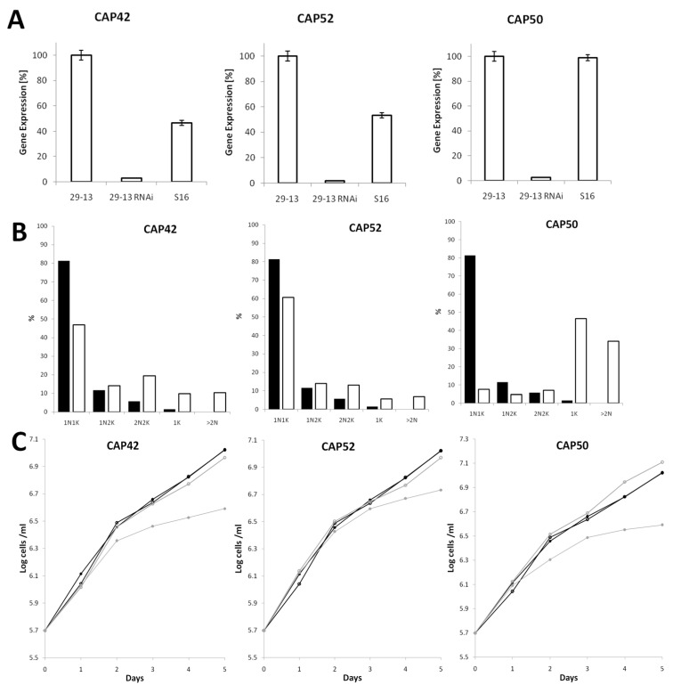
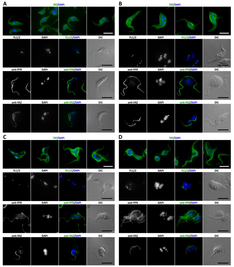
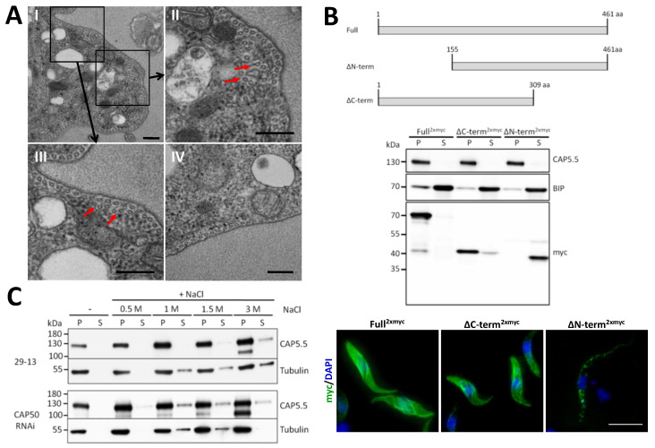
Trypanosome brucei, the causative agent of African sleeping sickness, harbours a highly ordered, subpellicular microtubule cytoskeleton that defines many aspects of morphology, motility and virulence. This array of microtubules is associated with a large number of proteins involved in its regulation. Employing proximity-dependent biotinylation assay (BioID) using the well characterised cytoskeleton-associated protein CAP5.5 as a probe, we identified CAP50 (Tb927.11.2610). This protein colocalises with the subpellicular cytoskeleton microtubules but not with the flagellum. Depletion by RNAi results in defects in cytokinesis, morphology and partial disorganisation of microtubule arrays. Published proteomics data indicate a possible association of CAP50 with two other, yet uncharacterised, cytoskeletal proteins, CAP52 (Tb927.6.5070) and CAP42 (Tb927.4.1300), which were therefore included in our analysis. We show that their depletion causes phenotypes similar to those described for CAP50 and that they are essential for cellular integrity.

Keywords: BioID; Trypanosoma brucei; cytoskeleton; mass spectrometry; microtubules.


Conflict of interest statement

The authors declare no conflict of interest.

Figures

Figure 1
Figure 1
A CAP5.5-BirA*expressing cell line to identify CAP5.5 proximal proteins by BioID. (A) Ectopic expression of CAP5.5-myc-BirA*-myc (here simplified termed as CAP5.5-BirA*2xmyc) in procyclic trypanosomes (PC449) was induced by 1 µg/mL doxycycline with an excess of biotin in the culture medium. Samples were taken after 1 and 2 days and fractionated in the soluble (S) and the pellet (P) fraction using 1% NP40. Biotinylated proteins were detected by HRP conjugated streptavidin. CAP5.5-myc-BirA*-myc was detected using an anti-myc antibody. Cells ectopically expressing CAP5.52xmyc were used as control. BIP and tubulin served as the loading control for the soluble and the insoluble fractions. (B) Localisation of the CAP5.5-myc-BirA*-myc protein over the cell body in whole cells (left image) and 1% NP40 extracted cells (right image). Cells were stained using an antibody against the myc-tag and DAPI to stain the DNA. Scale bar, 10 µm. (C) Proteins of the pellet fraction were solubilised and biotinylated proteins were purified by a Strep-Tactin® gravity flow column: 1, input, 2, flow through, 3, 4 wash fractions and 5, elution fractions pooled. The framed area of the gel, corresponding to the 60–70 kD mass range, was cut out and analysed by mass spectrometry.
Figure 2
Figure 2
Characterisation of three cytoskeleton associated proteins. (A) Dox-inducible ectopic expression of C-terminally myc-tagged CAP42, CAP52 and CAP50 in T. brucei. The localisation of the CAPs to the cytoskeleton but excluding the flagellum was analysed by labelling 1% NP40 treated cells with anti-myc (green) and anti-PFR antibody (L13D6, magenta) to mark the flagellum. Scale bar = 5 µm (B) Dox-induced cells were fractionated with 1% NP40 and the soluble (S) and pellet (P) fractions were analysed for the myc-tagged proteins. Fractions corresponding to equal cell numbers were loaded in all lanes (C) The cytoskeleton-containing pellet fractions from C were treated with increasing amounts of NaCl and the supernatants (S) and pellets (P) were analysed for the myc-tagged proteins. Tubulin (TAT), CAP5.5 and BIP served as the loading control for the insoluble and soluble fraction, respectively. Representative blots of three biological replicate experiments are shown.
Figure 3
Figure 3
RNAi depletion of CAPs leads to reduced cell division and abnormal karyotypes in procyclic cells. (A) Relative mRNA expression levels of the three CAPs in procyclic (29-13), bloodstream (S16) and their respective RNAi cell lines (29-13 RNAi) measured by RT-qPCR. The RNAi cell lines were induced for 1 day. PFRA transcript was used for normalisation. (B) Analysis of cell cycle progression. Nuclei and kinetoplasts were visualised by DAPI staining. The number of nuclei (N) and kinetoplasts (K) was counted for each cell (n > 300). Black bars represent non-induced and white bars induced cells. Induction was for 3 days. (C) Cumulative cell growth of CAPs RNAi cell lines (grey lines, open circles: −tet; grey lines, closed circles: +tet) compared to wild type cells (black lines, open circles: −tet; black lines, closed circles: +tet). Growth curve data are averages of three independent clones.
Figure 4
Figure 4
Immunofluorescence analysis of procyclic cells depleted of CAP52 (B) CAP42 (C) and CAP50 (D) by RNAi compared to wild-type 29-13 cells (A). Cells were doxycycline-induced for 3 days and fixed with methanol. TAT: anti-α-tubulin, YL1/2: anti-tyrosinated-α-tubulin (marker for basal bodies), PFR: anti-paraflagellar rod proteins A/C (L13D6), FAZ: anti-flagellum attachment zone (L3D2). DNA was stained with DAPI. Scale bar = 10 µm.
Figure 5
Figure 5
Ultrastructural and biochemical characterisation of CAP50. (A) TEM of transverse sections through procyclic cells depleted of CAP50 after RNAi induction for 3 days. Boxed areas in panel (I) are magnified in panels (II) and (III), showing examples of disordered microtubule arrays (some marked by red arrows). Panel (IV) shows a cross-section of a wild-type cell. Scale bar, 200 nm. (B) Truncation constructs of CAP50 used to analyse contribution of the C- or the N-terminus to cytoskeleton association of the protein. Truncation constructs and the full-length protein as control were C-terminally 2xmyc-tagged. For Western blot analysis, induced cells were fractionated with 1% NP40 in pellet (P) and supernatant (S) and the distribution of the protein was detected with an anti-myc antibody. Anti-BIP and anti-CAP5.5 were used as loading controls for the soluble and insoluble cytoskeleton fractions, respectively. For immunofluorescence cytoskeletons were labelled with an anti-myc antibody and DAPI. Scale bar, 10 µm (C) Analysis of the impact of CAP50 depletion on CAP5.5 cytoskeleton association. CAP50 RNAi cells were induced for 3 days and extracted with 1% NP40. The cytoskeletal pellet fraction was subjected to increasing concentrations of NaCl and supernatant (S) and pellet (P) fractions were analysed for their CAP5.5 content by Western blotting. Anti-tubulin was used to track the integrity of the microtubules. Wild type cells (29-13) were used as a control. Equal cell number equivalents were loaded in each lane. Representative blots of three biological replicate experiments are shown.

Similar articles

Cited by

References

    1. Jékely G. Origin and evolution of the self-organizing cytoskeleton in the network of eukaryotic organelles. Cold Spring Harb. Perspect. Biol. 2014;6:a016030. doi: 10.1101/cshperspect.a016030. - DOI - PMC - PubMed
    1. Dawson S.C., Paredez A.R. Alternative cytoskeletal landscapes: Cytoskeletal novelty and evolution in basal excavate protists. Curr. Opin. Cell Biol. 2013;25:134–141. doi: 10.1016/j.ceb.2012.11.005. - DOI - PMC - PubMed
    1. Gull K. The cytoskeleton of trypanosomatid parasites. Annu. Rev. Microbiol. 1999;53:629–655. doi: 10.1146/annurev.micro.53.1.629. - DOI - PubMed
    1. Sinclair A.N., de Graffenried C.L. More than microtubules: The structure and function of the subpellicular array in trypanosomatids. Trends Parasitol. 2019;35:760–777. doi: 10.1016/j.pt.2019.07.008. - DOI - PMC - PubMed
    1. Hayes P., Varga V., Olego-Fernandez S., Sunter J., Ginger M.L., Gull K. Modulation of a cytoskeletal calpain-like protein induces major transitions in trypanosome morphology. J. Cell Biol. 2014;206:377–384. doi: 10.1083/jcb.201312067. - DOI - PMC - PubMed

LinkOut - more resources