. 2021 Oct 31;9(11):2271.
doi: 10.3390/microorganisms9112271.

Intestinal Dominance by Serratia marcescens and Serratia ureilytica among Neonates in the Setting of an Outbreak

Affiliations

Intestinal Dominance by Serratia marcescens and Serratia ureilytica among Neonates in the Setting of an Outbreak

Elias Dahdouh et al. Microorganisms. .

Abstract

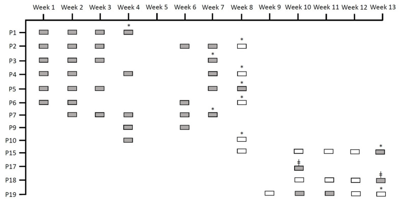
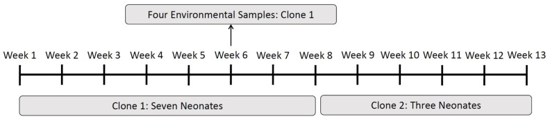
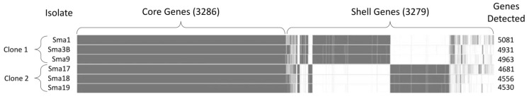
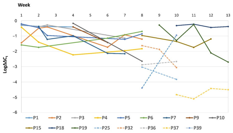
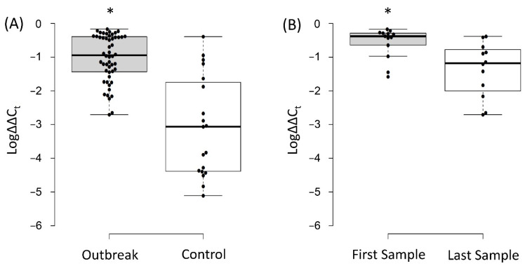
(1) Background: We determined the relevance of intestinal dominance by Serratia spp. during a neonatal outbreak over 13 weeks. (2) Methods: Rectal swabs (n = 110) were obtained from 42 neonates. Serratia spp. was cultured from swabs obtained from 13 neonates (Group 1), while the other 29 neonates were culture-negative (Group 2). Total DNA was extracted from rectal swabs, and quantitative PCRs (qPCRs) using Serratia- and 16SrRNA-gene-specific primers were performed. relative intestinal loads (RLs) were determined using ΔΔCt. Clonality was investigated by random amplified polymorphic DNA analysis and whole-genome sequencing. (3) Results: The outbreak was caused by Serratia marcescens during the first eight weeks and Serratia ureilytica during the remaining five weeks. Serratia spp. were detected by qPCR in all Group 1 neonates and eleven Group 2 neonates. RLs of Serratia spp. were higher in Group 1 as compared to Group 2 (6.31% vs. 0.09%, p < 0.05) and in the first swab compared to the last (26.9% vs. 4.37%, p < 0.05). Nine neonates had extraintestinal detection of Serratia spp.; eight of them were infected. RLs of the patients with extraintestinal spread were higher than the rest (2.79% vs. 0.29%, p < 0.05). (4) Conclusions: Intestinal dominance by Serratia spp. plays a role in outbreaks and extraintestinal spread.

Keywords: Serratia marcescens; Serratia ureilytica; intestinal dominance; neonatal ward; qPCR.


Conflict of interest statement

The authors declare no conflict of interest.

Figures

Figure 1
Figure 1
Rectal swabs obtained through routine epidemiological screening for Serratia spp. that were reused in our study. “P” stands for patient, grey rectangles indicate rectal swabs that were positive through routine culture, and white rectangles indicate rectal swabs that were negative through routine culture. All the rectangles depict samples that were included in our study. * Signifies that the patient was discharged, and ǂ signifies exitus.
Figure 2
Figure 2
Distribution of the two clones detected by random amplified polymorphic DNA analysis over the 13 weeks of the outbreak.
Figure 3
Figure 3
Pan-genome analysis for three isolates belonging to Clone 1 and three isolates belonging to Clone 2. The core genes are those common among all six isolates, while the shell genes are those that were different between the two clones.
Figure 4
Figure 4
Relative intestinal loads of Serratia spp. over time for all the patients that have had more than one episode of detection of this organism in rectal swabs. The dotted line indicates the relative loads for patients of Group 2 (i.e., samples obtained from culture-negative patients). Log∆∆Ct values of 0 represent 100% of the bacterial population, −1 is 10%, −2 is 1%, and so on until −6 (0.0001%), which is the detection limit.
Figure 5
Figure 5
(A) Log∆∆Ct values of Serratia spp. in samples obtained from Group 1 neonates (that had positive cultures) compared to those obtained from Group 2 neonates (culture-negative neonates). (B) Log∆∆Ct values of Serratia spp. in the first swab collected from each neonate of Group 1 compared to the last swab collected. Log∆∆Ct values of 0 represent 100% of the bacterial population, −1 is 10%, −2 is 1%, and so on until −6 (0.0001%), which is the detection limit. The “*” indicates statistical significance (p < 0.05).

References

    1. Mahlen S.D. Serratia infections: From military experiments to current practice. Clin. Microbiol. Rev. 2011;24:755–791. doi: 10.1128/CMR.00017-11. - DOI - PMC - PubMed
    1. Quinn L., Ailsworth M., Matthews E., Kellams A., Shirley D.A. Serratia marcescens Colonization Causing Pink Breast Milk and Pink Diapers: A Case Report and Literature Review. Breastfeed. Med. 2018;13:388–394. doi: 10.1089/bfm.2018.0002. - DOI - PubMed
    1. Ottolini K.M., Litke-Wager C.A., Johnson R.L., Schulz E.V. Serratia Chorioamnionitis and Culture Proven Sepsis in a Preterm Neonate: A Case Report and Review of the Literature. Pediatric Infect. Dis. J. 2020;40:e62–e65. doi: 10.1097/INF.0000000000002962. - DOI - PubMed
    1. The European Committee on Antimicrobial Susceptibility Testing (EUCAST) Expert Rules on Enterobacterales. Version 3.2. 2021. [(accessed on 19 October 2021)]. Available online: https://www.eucast.org/fileadmin/src/media/PDFs/EUCAST_files/Expert_Rule....
    1. Fisher M., Diago-Navarro E., Kaplun O., Sin E., Fries B.C. Serratia marcescens Strains Carrying blaKPC-2 and blaKPC-3 Carbapenemase Associated With Chronic Mechanical Ventilation. Open Forum Infect. Dis. 2018;5:S362. doi: 10.1093/ofid/ofy210.1029. - DOI

Grants and funding

LinkOut - more resources