. 2021 Nov 5;11(11):2974.
doi: 10.3390/nano11112974.

CD123-Targeted Nano-Curcumin Molecule Enhances Cytotoxic Efficacy in Leukemic Stem Cells

Affiliations

CD123-Targeted Nano-Curcumin Molecule Enhances Cytotoxic Efficacy in Leukemic Stem Cells

Wariya Nirachonkul et al. Nanomaterials (Basel). .

Abstract

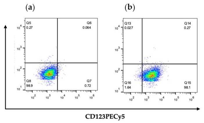
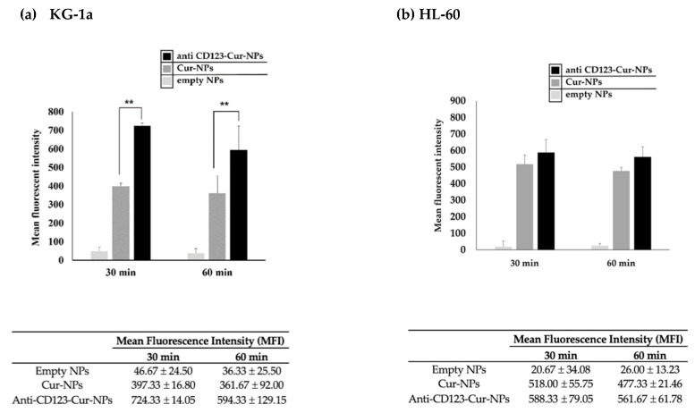
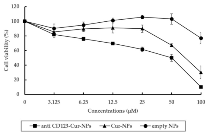
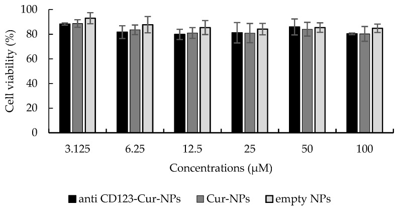
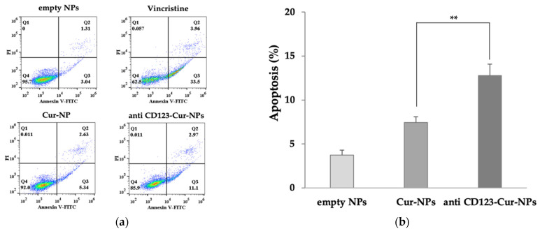
Acute myeloblastic leukemia (AML) is a disease with a high rate of relapse and drug resistance due to the remaining leukemic stem cells (LSCs). Therefore, LSCs are specific targets for the treatment of leukemia. CD123 is specifically expressed on LSCs and performs as a specific marker. Curcumin is the main active compound of a natural product with low toxicity for humans. It has been reported to inhibit leukemic cell growth. However, curcumin is practically insoluble in water and has low bioavailability. In this study, we aimed to formulate curcumin nanoparticles and conjugate with the anti-CD123 to overcome the low water solubility and improve the targeting of LSCs. The cytotoxicity of both curcumin-loaded PLGA/poloxamer nanoparticles (Cur-NPs) and anti-CD123-curcumin-loaded PLGA/poloxamer nanoparticles (anti-CD123-Cur-NPs) were examined in KG-1a cells. The results showed that Cur-NPs and Cur-NPs-CD123 exhibited cytotoxic effects on KG-1a cells with the IC50 values of 74.20 ± 6.71 and 41.45 ± 5.49 µM, respectively. Moreover, anti-CD123-Cur-NPs induced higher apoptosis than Cur-NPs. The higher uptake of anti-CD123-Cur-NPs in KG-1a cells was confirmed by using flow cytometry. In conclusion, the anti-CD123-Cur-NPs formulation improved curcumin's bioavailability and specific targeting of LSCs, suggesting that it is a promising drug delivery system for improving the therapeutic efficacy against AML.

Keywords: acute myeloblastic leukemia; anti-CD123; curcumin; leukemic stem cells; nanoparticles.


Conflict of interest statement

The authors declare no conflict of interest.

Figures

Figure 1
Figure 1
(a) FTIR spectra of the carboxylated poloxamer (top) and hydroxylated poloxamer (bottom); (b) FTIR spectrum of the anti-CD123-Cur-NP conjugate.
Figure 2
Figure 2
Characterization of nanoparticles. (a) Color of empty NPs, Cur-NPs, and anti-CD123-Cur-NPs. (b) Size measurement of the nanoparticles using dynamic light scattering (DLS); (b1) empty NPs, (b2) Cur-NPs, and (b3) anti-CD123-Cur-NPs. (c) TEM image of (c1) empty NPs, (c2) nontargeted Cur-NPs, and (c3) anti-CD123-Cur-NPs.
Figure 3
Figure 3
Surface expression of CD123 on KG-1a cells. Cells were stained with (a) isotype-matched control antibody or (b) CD123PECy5 antibody. Stained cells were assessed through flow cytometry.
Figure 4
Figure 4
Cellular uptake of anti-CD123-Cur-NPs or nontargeted Cur-NPs in KG-1a and HL-60 leukemia cell lines. KG-1a (CD123+) (a) and HL-60 (CD123-) (b) cells were incubated with anti-CD123-Cur-NPs or Cur-NPs for 30 and 60 min, and fluorescent intensity was determined through flow cytometry. The experimental data were collected in three independent experiments, and the results were determined using one-way ANOVA. The statistical differences were considered significant at ** p < 0.01.
Figure 5
Figure 5
Cytotoxicity of NPs in KG-1a cells. Cell viability of KG-1a cells was assessed with an MTT assay. The data are shown as mean ± SD from three independent experiments.
Figure 6
Figure 6
Cytotoxicity of NPs to peripheral blood mononuclear cells (PBMCs). PBMCs were treated with anti-CD123-Cur-NPs, Cur-NPs, and empty NPs for 48 h. The MTT assay determined the cell viability. Each bar represents the mean ± SD of three independent experiments performed in triplicate.
Figure 7
Figure 7
Apoptosis assay using flow cytometry after staining with double annexin V-FITC/propidium iodide (PI). KG-1a cells were treated with empty NPs, Cur-NPs, and anti-CD123-Cur-NPs. (a) Representative flow cytometry dot plot indicating the cell population in apoptotic and necrotic quadrants after treatment with different NPs. (b) The percentage of apoptotic cells was statistically compared. The data are shown as mean ± SD from three independent experiments. ** p < 0.05 between groups.

References

    1. Ladikou E.E., Sivaloganathan H., Pepper A., Chevassut T. Acute myeloid leukaemia in its niche: The bone marrow microenvironment in acute myeloid leukaemia. Curr. Oncol. Rep. 2020;22:27. doi: 10.1007/s11912-020-0885-0. - DOI - PMC - PubMed
    1. Döhner H., Weisdorf D.J., Bloomfield C.D. Acute myeloid leukemia. N. Engl. J. Med. 2015;373:1136–1152. doi: 10.1056/NEJMra1406184. - DOI - PubMed
    1. Yilmaz M., Wang F., Loghavi S., Bueso-Ramos C., Gumbs C., Little L., Song X., Zhang J., Kadia T., Borthakur G., et al. Late relapse in acute myeloid leukemia (AML): Clonal evolution or therapy-related leukemia. Blood Cancer J. 2019;9:7. doi: 10.1038/s41408-019-0170-3. - DOI - PMC - PubMed
    1. Döhner H., Estey E.H., Amadori S., Appelbaum F.R., Büchner T., Burnett A.K., Dombret H., Fenaux P., Grimwade D., Larson R.A., et al. Diagnosis and management of acute myeloid leukemia in adults: Recommendations from an international expert panel, on behalf of the european leukemia. Blood. 2010;115:453–474. doi: 10.1182/blood-2009-07-235358. - DOI - PubMed
    1. Jonas B.A. On the origin of relapse in AML. Sci. Transl. Med. 2017;9:eaan8205. doi: 10.1126/scitranslmed.aan8205. - DOI - PMC - PubMed

LinkOut - more resources