

Silver-Protein Nanocomposites as Antimicrobial Agents
- PMID: 34835774
- PMCID: PMC8617916
- DOI: 10.3390/nano11113006
Silver-Protein Nanocomposites as Antimicrobial Agents
Abstract
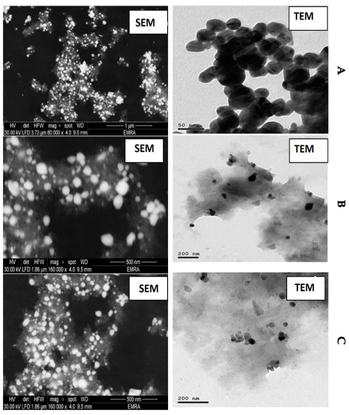
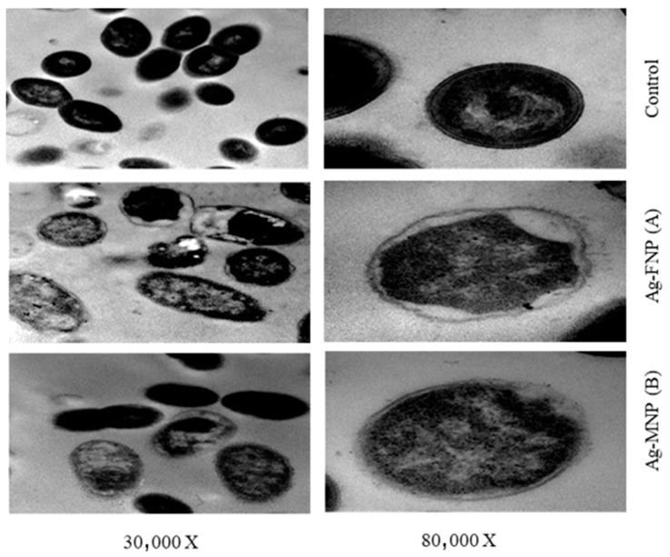
The use of nanomaterials alone or in composites with proteins is a promising alternative to inhibit pathogenic bacteria. In this regard, this study used seed proteins from both fenugreek (Trigonella foenum-graecum L.) (FNP) and mung bean (Viga radiate) (MNP), with silver nanoparticles (Ag-NPs) and nanocomposites of either Ag-NPs plus FNP (Ag-FNP) or Ag-NPs plus MNP (Ag-MNP) as inhibitory agents against pathogenic bacteria. FNP and MNP were isolated from fenugreek seeds and mung bean seeds, respectively, and fractionated using Sodium Dodecyl Sulfate-Polyacrylamide Gel Electrophoresis (SDS-PAGE). Both FNP and MNP were immobilized with Ag-NPs to synthesize the nanocomposites Ag-FNP and Ag-MNP, respectively. The physicochemical characteristics of Ag-NPs and their composites with proteins were studied by X-ray Diffraction (XRD), dynamic light scattering (DLS), the zeta potential, Scanning and Transmission Electron Microscopy (SEM and TEM, respectively), Atomic Force Microscopy (AFM), and the Brunauer-Emmett-Teller isotherm (BET), elucidating their structural parameters, size distribution, size charges, size surface morphology, particle shape, dimensional forms of particles, and specific surface area, respectively. The sole proteins, Ag-NPs, and their nanocomposites inhibited pathogenic Gram-positive and Gram-negative bacteria. The inhibitory activities of both nanocomposites (Ag-FNP and Ag-MNP) were more than those obtained by either Ag-NPs or proteins (FNP, MNP). Minimum inhibitory concentrations (MICs) of Ag-FNP were very low (20 and 10 µg mL-1) against Salmonellatyphimurium and Pseudomonasaerugenosa, respectively, but higher (162 µg mL-1) against E. coli and Listeriamonocytogenes. MICs of Ag-MNP were also very low (20 µg mL-1) against Staphylococcusaureus but higher (325 µg mL-1) against Listeriamonocytogenes. TEM images of Staphylococcusaureus and Salmonellatyphimurium, treated with Ag-FNP and Ag-MNP, at their MIC values, showed asymmetric, wrinkled exterior surfaces, cell deformations, cell depressions, and diminished cell numbers.
Keywords: antibacterial activity; fenugreek; mung bean; nanocomposite; seed proteins; silver nanoparticles.
Conflict of interest statement
The authors declare no conflict of interest.
Figures


References
-
- Askoura M., Saed N., Enan G., Askora A. Characterization of polyvalent bacteriophages targeting multi drug resistant Klebsiella pneumonia with enhanced anti -biofilm activity. Appl. Biochem. Microbiol. 2021;57:117–126. doi: 10.1134/S000368382101004X. - DOI
-
- Ismaiel A.A., Ali A.E., Enan G. Incidence of Listeria in Egyptian Meat and Dairy Samples . Food Sci. Biotechnol. 2014;23:179–185.
-
- Abdel-Shafi S., Ouda S.M., El-Balate I., Enan G. Characterization and identification of multidrug resistant bacteria from some Egyptian patients. Biotechnology. 2013;12:65–73. doi: 10.3923/biotech.2013.65.73. - DOI
-
- El-Sayed T.I., Atef D., Amer M., Mahdy A., Enan G. Molecular characterization and inhibition by natural agents of multidrug resistant Candida strains causing vaginal candidiasis. Res. J. Med. 2015;9:1–7.
LinkOut - more resources
Full Text Sources
Molecular Biology Databases
Miscellaneous
