. 2021 Nov 10;11(11):3017.
doi: 10.3390/nano11113017.

In Vitro and In Vivo Biocompatibility of Boron/Nitrogen Co-Doped Carbon Nano-Onions

Affiliations

In Vitro and In Vivo Biocompatibility of Boron/Nitrogen Co-Doped Carbon Nano-Onions

Marta d'Amora et al. Nanomaterials (Basel). .

Abstract

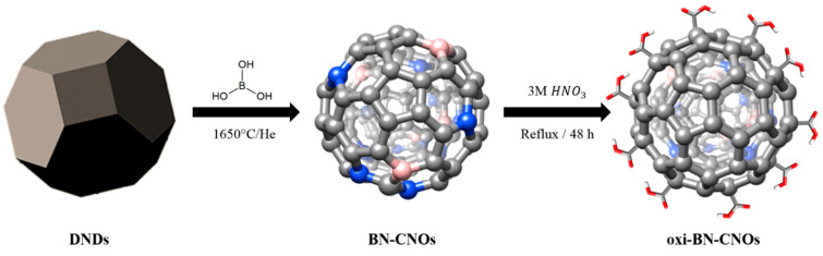
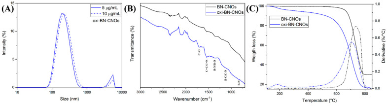
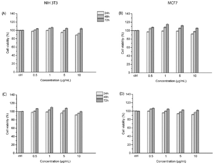
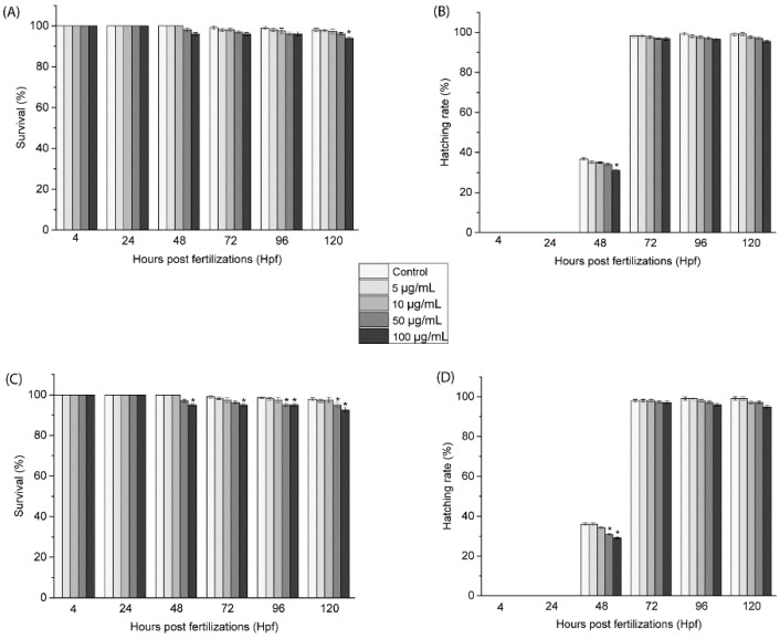
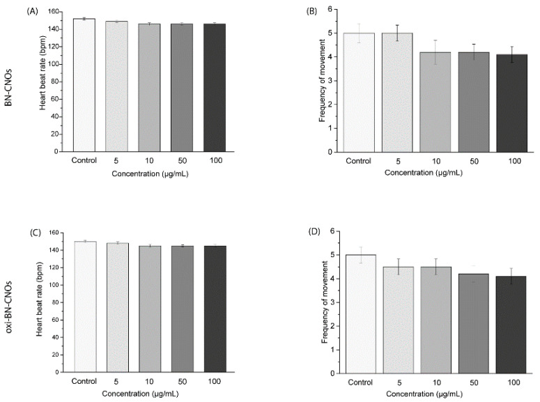
Boron/nitrogen, co-doped, carbon nano-onions (BN-CNOs) have recently shown great promise as catalysts for the oxygen reduction reaction, due to the improved electronic properties imparted by the dopant atoms; however, the interactions of BN-CNOs with biological systems have not yet been explored. In this study, we examined the toxicological profiles of BN-CNOs and oxidized BN-CNOs (oxi-BN-CNOs) in vitro in both healthy and cancer cell lines, as well as on the embryonic stages of zebrafish (Danio rerio) in vivo. The cell viabilities of both cell lines cells were not affected after treatment with different concentrations of both doped CNO derivatives. On the other hand, the analysis of BN-CNOs and oxidized BN-CNO interactions with zebrafish embryos did not report any kind of perturbations, in agreement with the in vitro results. Our results show that both doped CNO derivatives possess a high biocompatibility and biosafety in cells and more complex systems.

Keywords: biosafety; carbon nano-onion; cells; heteroatom doping; zebrafish.


Conflict of interest statement

The authors declare no conflict of interest.

Figures

Figure 1
Figure 1
Synthetic procedure for the production of BN-CNOs and oxi-BN-CNOs.
Figure 2
Figure 2
HRTEM images of BN-CNOs (A) and oxi-BN-CNOs (C), showing polyhedral-shaped CNOs. EELS spectra and corresponding STEM images as insets of BN-CNOs (B) and oxi-BN-CNOs (D), showing distinct B, C, and N–K edges. Blue and red lines in panel D refer to the external and internal layers, as depicted in the inset.
Figure 3
Figure 3
XPS survey (A,E) and high-resolution C 1s (B,F), B 1s (C,G), and N 1s (D,H) XPS spectra of BN-CNOs (upper panel) and oxi-BN-CNOs (lower panel), including peak deconvolution and experimental (black) and fitting curves (red).
Figure 4
Figure 4
UV-Vis absorption spectra of (A) BN- and (B) oxi-BN-CNO dispersions in deionized water at different concentrations (5, 10, 20, 50 and 100 µg/mL). (C) Pictures of the solutions at the different concentrations.
Figure 5
Figure 5
(A) DLS spectra of oxi-BN-CNO dispersions in deionized water at concentrations of 5 and 10 μg/mL. (B) FTIR spectra and (C) TGA (solid lines) and corresponding weight loss derivatives (dotted lines) of BN—(black) and oxi-BN-CNOs (blue).
Figure 6
Figure 6
Cellular viability of NIH 3T3 and MCF7 cells treated with various concentrations of BN- (A,B) and oxi-BN-CNOs (C,D) for 24, 48, and 72 h. Data are expressed as mean ± standard deviations of three replicates.
Figure 7
Figure 7
Survival rates (AC) and hatching rates (BD) of embryos exposed to different concentrations of BN- and oxi-BN-CNOs (5, 10, 50, and 100 μg/mL). Data are calculated as means ± S.D., from three independent experiments, n = 80 (* indicate p ≤ 0.01 compared to the control).
Figure 8
Figure 8
Heart rate (AC) and frequency of voluntary movements (BD) of larvae at 72 hpf exposed to different concentrations of BN- and oxi-BN-CNOs.

Similar articles

Cited by

References

    1. Ugarte D. Curling and closure of graphitic networks under electron-beam irradiation. Nature. 1992;359:707–709. doi: 10.1038/359707a0. - DOI - PubMed
    1. Camisasca A., Giordani S. Carbon nano-onions in biomedical applications: Promising theranostic agents. Inorg. Chim. Acta. 2017;468:67–76. doi: 10.1016/j.ica.2017.06.009. - DOI
    1. Lettieri S., Camisasca A., d’Amora M., Diaspro A., Uchida T., Nakajima Y., Yanagisawa K., Maekawa T., Giordani S. Far-red fluorescent carbon nano-onions as a biocompatible platform for cellular imaging. RSC Adv. 2017;7:45676–45681. doi: 10.1039/C7RA09442F. - DOI
    1. d’Amora M., Maffeis V., Brescia R., Barnes D., Scanlan E., Giordani S. Carbon Nano-Onions as Non-Cytotoxic Carriers for Cellular Uptake of Glycopeptides and Proteins. Nanomaterials. 2019;9:1069. doi: 10.3390/nano9081069. - DOI - PMC - PubMed
    1. d’Amora M., Camisasca A., Lettieri S., Giordani S. Toxicity Assessment of Carbon Nanomaterials in Zebrafish during Development. Nanomaterials. 2017;7:414. doi: 10.3390/nano7120414. - DOI - PMC - PubMed

LinkOut - more resources