. 2021 Oct 30;13(11):3904.
doi: 10.3390/nu13113904.

Anti-Osteoporosis Effects of the Eleutherococcus senticosus, Achyranthes japonica, and Atractylodes japonica Mixed Extract Fermented with Nuruk

Affiliations

Anti-Osteoporosis Effects of the Eleutherococcus senticosus, Achyranthes japonica, and Atractylodes japonica Mixed Extract Fermented with Nuruk

So Young Eun et al. Nutrients. .

Abstract

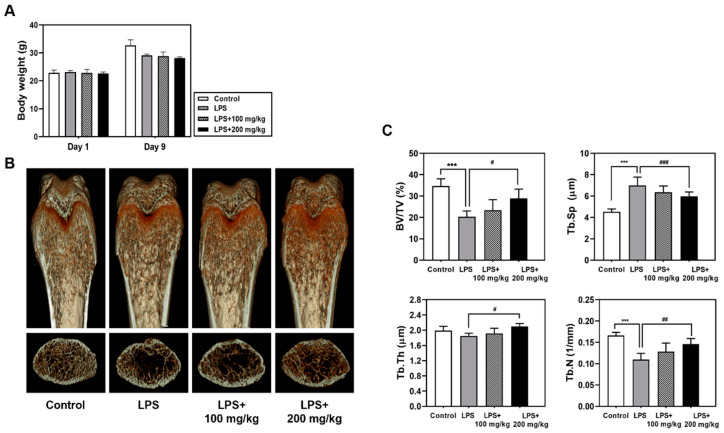
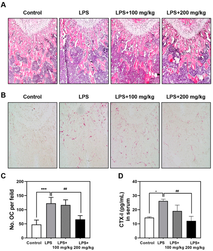
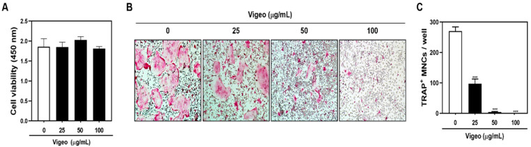
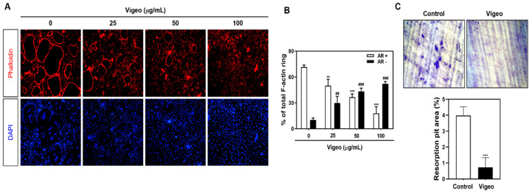
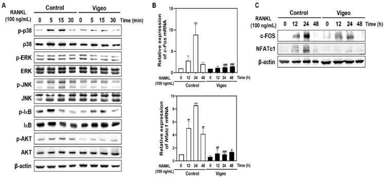
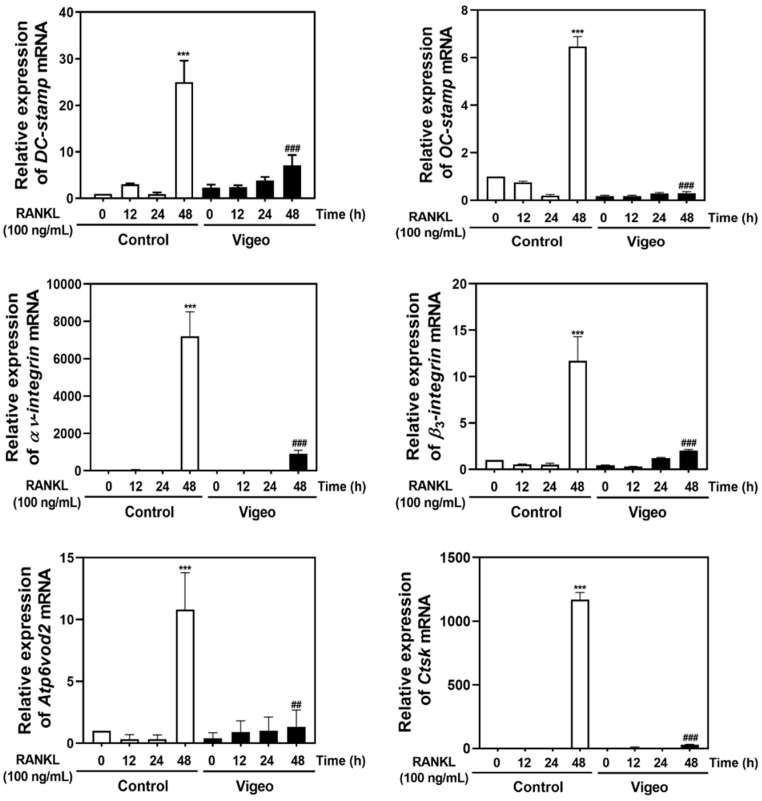
Vigeo is a mixture of fermented extracts of Eleutherococcus senticosus Maxim (ESM), Achyranthes japonica (Miq.) Nakai (AJN), and Atractylodes japonica Koidzumi (AJK) manufactured using the traditional Korean nuruk fermentation method. Although the bioactive effects of ESM, AJN, and AJK have already been reported, the pharmacological effects of Vigeo have not been proven. Therefore, in this study, we investigated whether Vigeo had inhivitory effects on lipopolysaccharide (LPS)-induced inflammatory bone loss in vivo and receptor activator of nuclear factor-B ligand (RANKL)-induced osteoclastogenesis and the related mechanism in vitro. Vigeo administration conferred effective protection against bone loss induced by excessive inflammatory response and activity of osteoclasts in LPS-induced inflammatory osteoporosis mouse model. In addition, Vigeo significantly suppressed the formation of tartrate-resistant acid phosphatase-positive osteoclasts induced by RANKL and inhibited F-actin formation and bone resorbing activity without any cytotoxicity. Moreover, Vigeo significantly inhibited RANKL-induced phosphorylation of p38, ERK, JNK, IκB, and AKT and degradation of IkB. Additionally, Vigeo strongly inhibited the mRNA and protein expression of c-FOS and NFATc1 and subsequently attenuated the expression of osteoclast specific marker genes induced by RANKL. We demonstrated for the first time the anti-osteoporosis effect of Vigeo, suggesting that it could be a potential therapeutic candidate for the treatment of osteoclast-mediated inflammatory bone diseases.

Keywords: Vigeo; bone resorption; nuruk fermentation; osteoclasts; osteoporosis.


Conflict of interest statement

The authors declare no conflict of interest.

Figures

Figure 1
Figure 1
Vigeo attenuates lipopolysaccharide (LPS)-induced inflammatory bone loss. Mice were intraperitoneally injected with LPS (10 mg/kg) followed by treatment with indicated concentrations of Vigeo for 7 days. (A) Body weight measurement during the experimental period. (B) Representative three-dimensional micro-computed tomography (micro-CT) images of the coronal and transverse sections in the distal femurs of a mouse from each group. (C) Measurement of the trabecular morphometric parameters of bone volume/total volume (BV/TV), trabecular separation (Tb. Sp), trabecular thickness (Tb. Th), and trabecular number (Tb. N) in the proximal femurs using the micro-CT data using the INFINITT-Xelis software 1.16. Statistical analysis was performed using one-way analysis of variance (ANOVA) followed by Tukey’s posthoc test for comparisons among three groups. *** p < 0.01 versus the control group; # p < 0.05, ## p < 0.01, ### p < 0.001 versus the LPS group.
Figure 2
Figure 2
Vigeo inhibits lipopolysaccharide (LPS)-induced osteoclast activation in mice. (A) LPS injected mice were sacrificed after 9 days, and dissected femurs were fixed, decalcified, paraffin-embedded, and sectioned. The slices were stained with hematoxylin and eosin (H&E). (B) Slices stained with tartrate-resistant acid phosphatase (TRAP). (C) The number of osteoclasts per visual field of tissue was measured by histomorphometric analysis. (D) Blood samples were collected from mice and centrifuged to obtain serum for the evaluation of CTX-I levels by ELISA. Statistical analysis was performed using one-way analysis of variance (ANOVA) followed by Tukey’s posthoc test for comparisons among the three groups. * p < 0.05, *** p < 0.001 versus the control group; ## p < 0.01 versus the LPS group.
Figure 3
Figure 3
Vigeo attenuates receptor activator of nuclear factor-B ligand (RANKL)-induced osteoclast differentiation without cytotoxicity. (A) Bone marrow macrophages (BMMs) were cultured with 30 ng/mL M-CSF and the indicated concentration of Vigeo for 3 days. Cell viability of Sec was evaluated using the XTT assay by measuring absorbance at 450 nm. (B) BMMs were seeded with M-CSF (30 ng/mL) and stimulated with RANKL (100 ng/mL) and the indicated concentrations of Vigeo for 3 days. TRAP staining was performed to assess osteoclast formation and representative images were captured. (C) TRAP-positive multinucleated cells counted as osteoclasts (nuclei > 5). *** p < 0.001 versus the control group.
Figure 4
Figure 4
Vigeo suppresses the F-actin ring formation and bone-resorbing activity of mature osteoclasts. (A) Bone marrow macrophages (BMMs)were cultured with M-CSF (30 ng/mL) and receptor activator of nuclear factor-B ligand (RANKL) (100 ng/mL) in the presence of the indicated concentrations of Vigeo. The cells were fixed, permeabilized, and stained with phalloidin and 4′,6-diamidino-2-phenylindole (DAPI). The cells were examined under a confocal laser scanning microscope. (B) Normal actin ring (AR+) osteoclasts and disrupted actin ring (AR−) osteoclasts were counted and compared with the total number of osteoclasts that contain three or more nuclei. The graph shows the comparison between relative percentage of osteoclasts that express AR+ and AR−. ** p < 0.01 and *** p < 0.001 versus AR+ osteoclasts in control group; ## p < 0.01 and ### p < 0.001 versus AR− osteoclasts in control group. (C) Mature osteoclasts from the co-culture system were seeded in dentin slices for 48 h with or without Vigeo (100 μg/mL). Cells attached to the hydroxyapatite-coated plate were removed, and the plates were photographed under a light microscope. *** p < 0.001 versus the control.
Figure 5
Figure 5
Vigeo affects receptor activator of nuclear factor-B ligand (RANKL)-induced phosphorylation of early signals and expression of c-FOS and NFATc1. (A) Bone marrow macrophages (BMMs)were seeded in the presence of M-CSF (10 ng/mL) for 1 day and then starved for 4 h. (A) Vigeo was used to pretreat the cells for 1 h followed by 0, 5, 15, and 30 min of RANKL stimulation. Whole-cell proteins were extracted by lysis using RIPA buffer and the protein expression was detected by western blotting using the indicated antibodies. (B) BMMs were cultured with M-CSF (30 ng/mL) and RANKL (100 ng/mL) in the presence or absence of Vigeo (100 μg/mL). Total RNA was isolated from the cells and mRNA expression levels of c-Fos and Nfatc1 were evaluated by real-time RT-PCR. ** p < 0.01, *** p < 0.001 versus the control; # p < 0.05, ## p < 0.01, ### p < 0.001 versus the control at indicated times. (C) BMMs were cultured with M-CSF (30 ng/mL) and RANKL (100 ng/mL) in the presence or absence of Vigeo (100 μg/mL). Protein expression was detected using western blotting with the indicated antibodies. β-actin was used as an internal control.
Figure 6
Figure 6
Vigeo inhibits receptor activator of nuclear factor-B ligand (RANKL)-induced mRNA expression of DC-stamp, OC-stamp, αv-integrin, β3-integrin, Atp6v0d2, and CtsK. Bone marrow macrophages (BMMs)were incubated with M-CSF (30 ng/mL) and RANKL (100 ng/mL) in the presence or absence of Vigeo (100 μg/mL) for the indicated time. Total RNA was isolated from cells using TRIzol reagent, and the mRNA expression of DC-stamp, OC-stamp, αv-integrin, β3-integrin, Atp6v0d2, and CtsK was analyzed by real-time RT-PCR. *** p < 0.001 versus the control at 0 h; ## p < 0.01, ### p < 0.001 versus the control at 48 h.

References

    1. Kanis J.A., Melton L.J., Christiansen C., Johnston C.C., Khaltaev N. The diagnosis of osteoporosis. J. Bone Miner. Res. 1994;9:1137–1141. doi: 10.1002/jbmr.5650090802. - DOI - PubMed
    1. Rousseau S., Kyomugasho C., Celus M., Hendrickx M.E.G., Grauwet T. Barriers impairing mineral bioaccessibility and bioavailability in plant-based foods and the perspectives for food processing. Crit. Rev. Food Sci. Nutr. 2020;60:826–843. doi: 10.1080/10408398.2018.1552243. - DOI - PubMed
    1. Xutian S., Zhang J., Louise W. New exploration and understanding of traditional Chinese medicine. Am. J. Chin. Med. 2009;37:411–426. doi: 10.1142/S0192415X09006941. - DOI - PubMed
    1. Cheon Y.H., Baek J.M., Park S.H., Ahn S.J., Lee M.S., Oh J.M., Kim J.Y. Stauntonia hexaphylla (Lardizabalaceae) leaf methanol extract inhibits osteoclastogenesis and bone resorption activity via proteasome-mediated degradation of c-Fos protein and suppression of NFATc1 expression. BMC Complementary Altern. Med. 2015;15:280. doi: 10.1186/s12906-015-0801-6. - DOI - PMC - PubMed
    1. Lim D.W., Kim J.G., Lee Y.S., Cha S.H., Kim Y.T. Preventive Effects of Eleutherococcus senticosus Bark extract in OVX-induced osteoporosis in rats. Molecules. 2013;18:7998–8008. doi: 10.3390/molecules18077998. - DOI - PMC - PubMed