

Medial patellofemoral ligament reconstruction combined with biplanar supracondylar femoral derotation osteotomy in recurrent patellar dislocation with increased femoral internal torsion and genu valgum: a retrospective pilot study
- PMID: 34836529
- PMCID: PMC8626929
- DOI: 10.1186/s12891-021-04816-2
Medial patellofemoral ligament reconstruction combined with biplanar supracondylar femoral derotation osteotomy in recurrent patellar dislocation with increased femoral internal torsion and genu valgum: a retrospective pilot study
Abstract
Background: The purpose of this study was to evaluate the clinical and radiographic outcomes after medial patellofemoral ligament (MPFL) reconstruction combined with supracondylar biplanar femoral derotation osteotomy (FDO) in recurrent patellar dislocation (RPD) with increased femoral anteversion angle (FAA) and genu valgum.
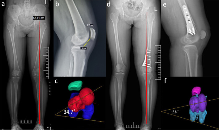
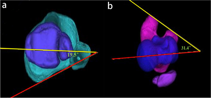
Methods: Between January 2017 to December 2020, a total of 13 consecutive patients (13 knees, 4 males and 9 females, mean age 18.7 (range, 15-29 years) with RPD with increased FAA (FAA > 25°) and genu valgum (mechanical axis deformity of ≥5°) who underwent supracondylar biplanar FDO using a Tomofix-locking plate combined with MPFL reconstruction in our institution were included. Preoperative full-leg standing radiographs, lateral views, and hip-knee-ankle computed tomography (CT) scans were used to evaluate the mechanical lateral distal femoral angle (mLDFA), anatomical femorotibial angle (aFTA), mechanical axis, patellar height, tibial tubercle-trochlear groove (TT-TG) distance, and torsional angle of the tibial and femoral in the axial plane. Patient reported outcomes were evaluated using the International Knee Documentation Committee (IKDC) score, Kujala score, Lysholm score, visual analog scale (VAS), and Tegner score preoperatively and postoperatively. Postoperative CT scans were used to evaluate the changes of FAA and TT-TG, and full-leg standing radiographs was used to evaluate the changes of mLDFA, aFTA, and mechanical axis.
Results: A total of 13 patients (13 knees) were included with an average follow-up period of 26.7 months (range 24-33). No cases developed wound infection, soft tissue irritation, and recurrent patellar dislocation during the follow-up period after surgery. Bone healing at the osteotomy site was achieved in all cases, and all patients regained full extension and flexion. Clinical outcomes (VAS, Kujala, IKDC, Lysholom, and Tegner scores) improved significantly at the final follow-up after surgery (p < 0.05). The mean mLDFA, aFTA, mechanical axis, and TT-TG distance showed statistically significant improvement following the combined surgery (p < 0.05), while the CDI did not change significantly after surgery (p>0.05).
Conclusion: MPFL reconstruction combined with supracondylar biplanar FDO showed satisfactory clinical outcomes and radiographic results in the short-term follow-up period.
Keywords: Alignment correction; Biplanar; Femoral anteversion angle; Femoral derotation osteotomy; Genu valgum; Medial patellofemoral ligament reconstruction; Recurrent patellar dislocation.
© 2021. The Author(s).
Conflict of interest statement
All the authors declare that they have no conflict of interest with any organization.
Figures
References
-
- Nelitz M, Dreyhaupt J, Williams SR, Dornacher D. Combined supracondylar femoral derotation osteotomy and patellofemoral ligament reconstruction for recurrent patellar dislocation and severe femoral anteversion syndrome: surgical technique and clinical outcome. Int Orthop. 2015;39(12):2355–2362. doi: 10.1007/s00264-015-2859-7. - DOI - PubMed
-
- Rogers BA. Patella alta: Association with patellofemoral alignment and changes in contact area during weight-bearing. J Bone Joint Surg Am. 2008;90(2):446–447. - PubMed
MeSH terms
LinkOut - more resources
Full Text Sources
Miscellaneous
