. 2021:79:89-132.
doi: 10.1016/bs.ampbs.2021.07.004. Epub 2021 Aug 13.

Extracellular haem utilization by the opportunistic pathogen Pseudomonas aeruginosa and its role in virulence and pathogenesis

Affiliations

Extracellular haem utilization by the opportunistic pathogen Pseudomonas aeruginosa and its role in virulence and pathogenesis

Susana Mouriño et al. Adv Microb Physiol. 2021.

Abstract

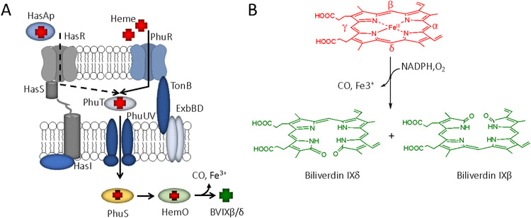
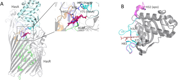
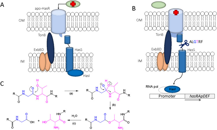
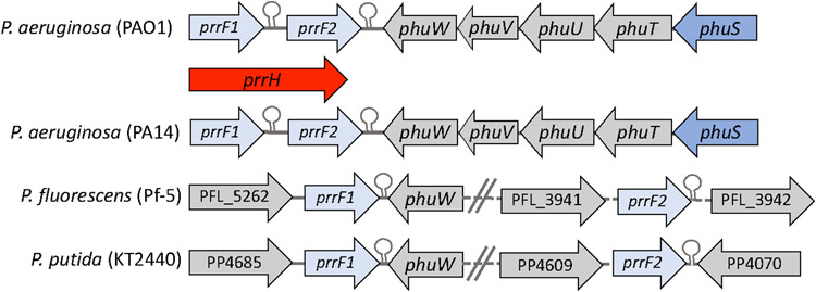
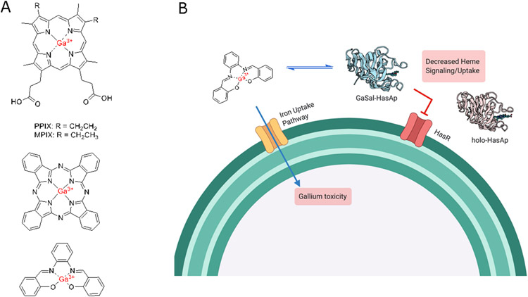
Iron is an essential micronutrient for all bacteria but presents a significant challenge given its limited bioavailability. Furthermore, iron's toxicity combined with the need to maintain iron levels within a narrow physiological range requires integrated systems to sense, regulate and transport a variety of iron complexes. Most bacteria encode systems to chelate and transport ferric iron (Fe3+) via siderophore receptor mediated uptake or via cytoplasmic energy dependent transport systems. Pathogenic bacteria have further lowered the barrier to iron acquisition by employing systems to utilize haem as a source of iron. Haem, a lipophilic and toxic molecule, presents a significant challenge for transport into the cell. As such pathogenic bacteria have evolved sophisticated cell surface signaling (CSS) and transport systems to sense and obtain haem from the host. Once internalized haem is cleaved by both oxidative and non-oxidative mechanisms to release iron. Herein we summarize our current understanding of the mechanism of haem sensing, uptake and utilization in Pseudomonas aeruginosa, its role in pathogenesis and virulence, and the potential of these systems as antimicrobial targets.

Keywords: Bacterial pathogenesis; Biliverdin; ECF σ-factor systems; Haem uptake and utilization; Iron and haem regulated sRNAs; Iron homeostasis; Pseudomonas aeruginosa; Transcriptional regulation.


Conflict of interest statement

Disclosure statement The authors are not aware of any affiliations, funding or financial interests that might be perceived as influencing the objectivity of this review.

Figures

Figure 1.
Figure 1.
Overview of P. aeruginosa haem uptake and utilization. A. Haem is bound by HasAp or extracted from hemoglobin (Hb) for transport across the outer membrane by the TonB-dependent receptors HasR and PhuR, respectively. In the periplasm haem is shuttled to the ABC transporter (PhuUV) by the periplasmic binding protein (PhuT) and actively transported across the cytoplasmic membrane by PhuUV. Haem is sequestered by the cytoplasmic haem binding protein (PhuS) for transfer to haem oxygenase (HemO). B. HemO oxidatively cleaves haem releasing iron, carbon monoxide (CO), and biliverdin IXβ/δ.
FIGURE 2.
FIGURE 2.. Structure of the HasA-HasR complex and HasAp.
(A) S. marcescens HasA docked to HasR. HasR, the membrane spanning β-barrel and extracellular loops are shown in grey, the N-terminal plug in green. The haemophore HasA is shown in cyan the haem in red and the HasR extended L7 and L8 loops that interact with HasA orange and blue, respectively. The boxed area is shown magnified in the right panel as a close-up view of haem ligated in HasR through H602 from the FRAP/PNPL L7 and H189 of the N-terminal plug. L8 I691 that provides the steric clash with the H32 loop of HasA shown in blue. The HasA protein is shown in cyan with the displaced H83 and Y75 as sticks. Adapted from PDB file 3CSL. (B) Overlay of the P. aeruginosa apo-HasAp and holo-HasAp structures. Apo- and holo HasAp H32 and Y75 loops shown in magenta and cyan, respectively. Overall fold shown in grey. Residues as labeled with the Y75-H83 hydrogen bond interaction highlighted in yellow. PDB files 3MOK (apo) and 3ELL holo).
FIGURE 3.
FIGURE 3.. Schematic representation of the ECF σ-factor signaling system.
(A) Signaling cascade in the “off” state prior to holo-HasAp binding. The N-terminal signaling domain as well as the TonB box are not bound by HasR. (B) Holo-HasA binds to HasR inducing a conformational change in the N-terminal signaling domain that recruits the anti-σ factor, promoting self-cleavage triggering the release of HasI. Additionally, it is thought that the TonB-box engages with the periplasmic TonB complex. (C) Proposed autocatalytic cleavage and corresponding N-O acyl rearrangement. The HasS self-cleavage motif ALGTRF located between the N and C-terminal domain with the catalytic residues shown in cyan (Thr) and blue (Gly). (C) (a) nucleophilic attack of Thr (cyan) on the preceding Gly residue (blue) forming a tetrahedral hydroxyoxazolidine intermediate. (b) rearrangement of the tetrahedral intermediate to yield the ester. (c) ester hydrolysis and peptide cleavage.
FIGURE 4.
FIGURE 4.. Genetic organization of the phu and prrF1,F2 locus in pathogenic and non-pathogenic Pseudomonas strains.
PrrH sRNA obtained on read through of the prrF1 Rho-independent terminator.
FIGURE 5.
FIGURE 5.. Proposed model for the heme-dependent modulation of PrrF and PrrH expression by PhuS.
(A) Equilibrium toward apo-PhuS leads to increase in the relative expression of PrrH on PhuS binding and reorganization of the prrF1 promoter . (B) Active haemuptake shifts the equilibrium toward holo-PhuS down regulating PrrH relative to PrrF1 and/or PrrF2. (C) Increased intracellular iron levels lead to Fur repression of the prrF1,2 operon. Adapted from Wilson et al, (2021).
FIGURE 6.
FIGURE 6.. Proposed mechanism for haem transfer from holo-HasAp to HasR.
(A) holo-HasAp WT interaction with HasR. A conformational change and the free energy gained on HasAp-HasR interaction drives the concerted release of the HasAp Y75 and H32 ligands treleasing haem to HasR. (B) holo-HasAp Y75A (or H32A) interaction with HasR. The open haem coordination site in Y75A (or H32A) mutant is occupied by a H2O molecule that on interaction with HasR is displaced forming the kinetically trapped intermediate leading to increased haem signaling and decreased transport. (C) holo-HasAp Y75H interaction with HasR. The free energy gained on protein-protein interaction of holo-HasAp Y75H with HasR is not sufficient to drive release of haem from holo-HasAp Y75H blocking both signaling and transport. Adapted from Dent et al, (2019).
FIGURE 7.
FIGURE 7.. Structure of PhuT and the Y. pestis HmuUV.
(A) holo-PhuT with β-sheet/loops in magenta and α-helices in cyan (PDB file 2R79). (B) Overall structural fold of the HmuUV transporter. HmuU membrane subunits are shown in blue and light blue, and the HmuV ATPase are subunits in orange and yellow. Bound phosphates shown in red (PDB file 4G1U).
FIGURE 8.
FIGURE 8.. Proposed mechanism of haem translocation by the haem ABC-transporters.
(A) Outward facing conformation where the TMD interactions form a periplasmic gate blocked from the cytoplasm. (B) Docking of the PBP releases haem to the channel causes the periplasmic gate to close giving the proposed occluded intermediate. (C) ATP hydrolysis drives a conformational change toward the inward facing conformation releasing haem to the cytoplasmic binding protein. (D) Binding of ATP releases the PBP and resets the transporter in the outward facing conformation.
FIGURE 9.
FIGURE 9.. Haem mimics as inhibitors of haem uptake and utilization.
(A) Gallium compounds including Ga3+-PPIX/MPIX (Top), Ga3+-Phthalocyanine (Middle) and Ga3+-Salophen (Bottom). (B) Schematic of Ga3+-Salophen as a substrate for active siderophore uptake and inhibition of the Has CSS cascade.

Similar articles

Cited by

References

    1. Afonina G, Leduc I, Nepluev I, Jeter C, Routh P, Almond G, et al. (2006). Immunization with the Haemophilus ducreyi hemoglobin receptor HgbA protects against infection in the swine model of chancroid. Infect Immun, 74(4), 2224–2232. - PMC - PubMed
    1. Agnoli K, Haldipurkar SS, Tang Y, Butt AT, & Thomas MS (2019). Distinct Modes of Promoter Recognition by Two Iron Starvation σ Factors with Overlapping Promoter Specificities. J Bacteriol, 201(3), e00507–00518. - PMC - PubMed
    1. Ainsaar K, Tamman H, Kasvandik S, Tenson T, & Horak R (2019). The TonBm-PocAB System Is Required for Maintenance of Membrane Integrity and Polar Position of Flagella in Pseudomonas putida. J Bacteriol, 201(17) e00303–00319. - PMC - PubMed
    1. Alontaga AY, Rodriguez JC, Schonbrunn E, Becker A, Funke T, Yukl ET, et al. (2009). Structural characterization of the hemophore HasAp from Pseudomonas aeruginosa: NMR spectroscopy reveals protein-protein interactions between Holo-HasAp and hemoglobin. Biochemistry, 48(1), 96–109. - PMC - PubMed
    1. Arivett BA, Fiester SE, Ohneck EJ, Penwell WF, Kaufman CM, Relich RF, et al. (2015). Antimicrobial Activity of Gallium Protoporphyrin IX against Acinetobacter baumannii Strains Displaying Different Antibiotic Resistance Phenotypes. Antimicrob Agents Chemother, 59(12), 7657–7665. - PMC - PubMed

Publication types