Review
. 2022 May;32(5):421-432.
doi: 10.1016/j.tcb.2021.10.006. Epub 2021 Nov 23.

Nucleation, stabilization, and disassembly of branched actin networks

Affiliations
Review

Nucleation, stabilization, and disassembly of branched actin networks

Alexis M Gautreau et al. Trends Cell Biol. 2022 May.

Abstract

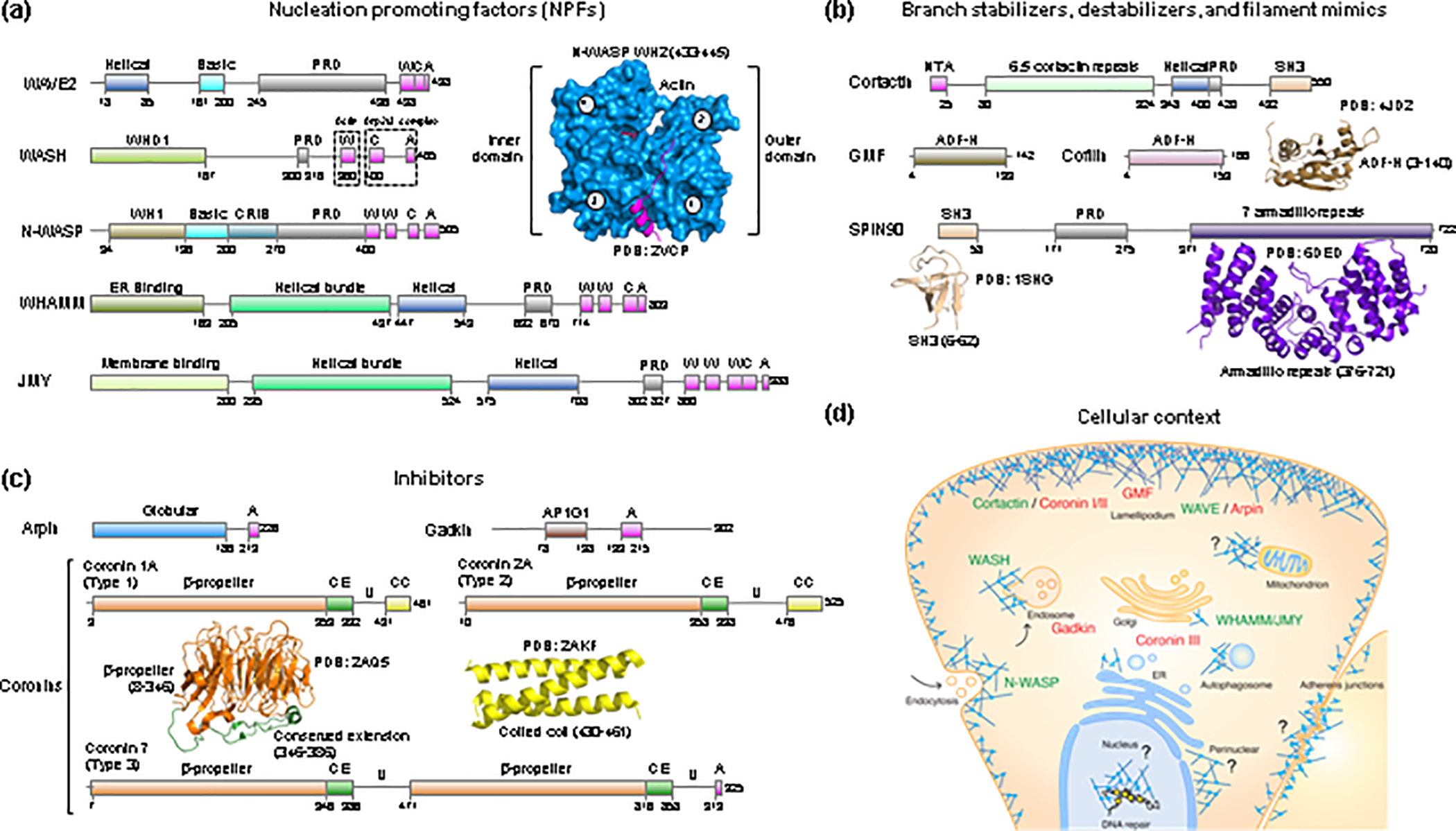
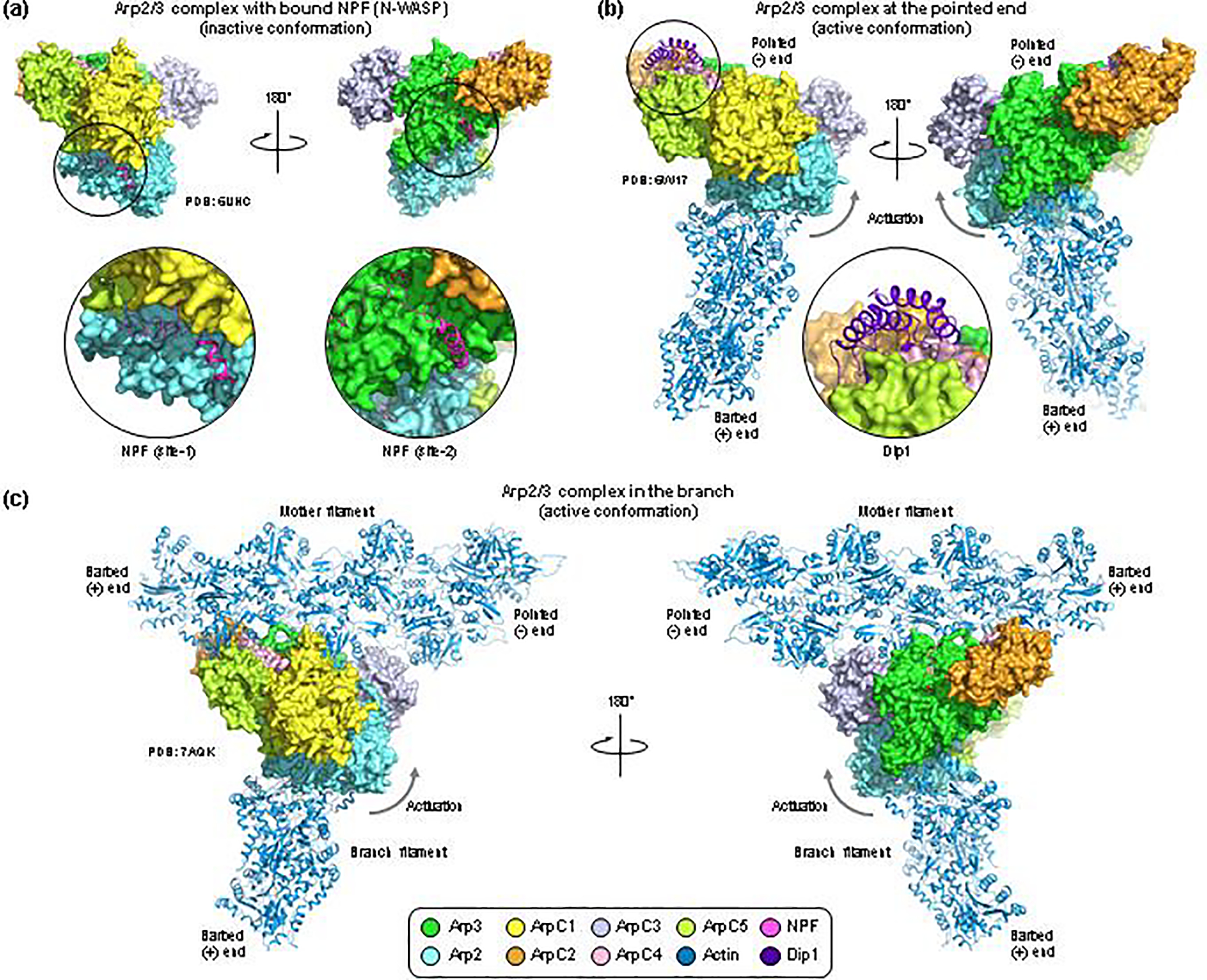
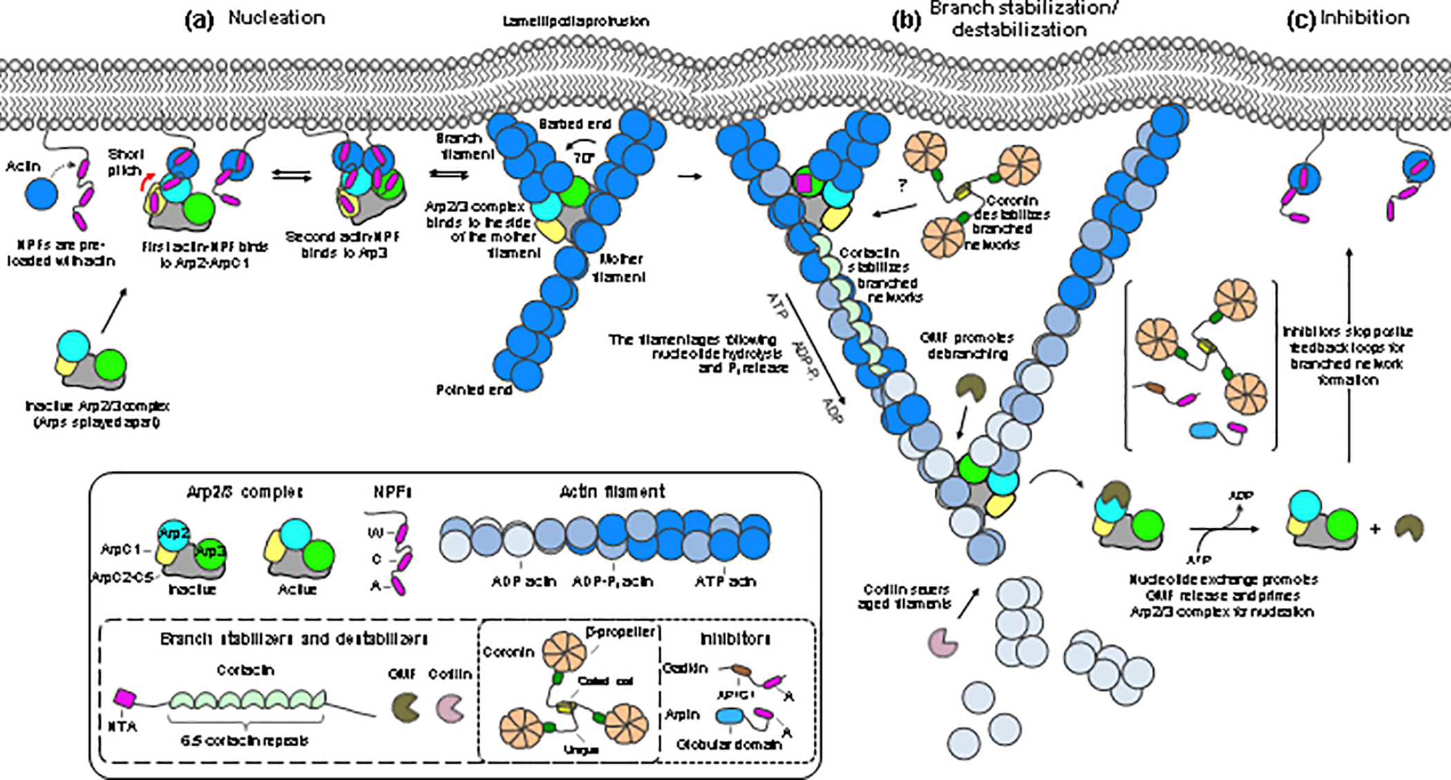
Arp2/3 complex is an actin filament nucleation and branching machinery conserved in all eukaryotes from yeast to human. Arp2/3 complex branched networks generate pushing forces that drive cellular processes ranging from membrane remodeling to cell and organelle motility. Several molecules regulate these processes by directly inhibiting or activating Arp2/3 complex and by stabilizing or disassembling branched networks. Here, we review recent advances in our understanding of Arp2/3 complex regulation, including high-resolution cryoelectron microscopy (cryo-EM) structures that illuminate the mechanisms of Arp2/3 complex activation and branch formation, and novel cellular pathways of branch formation, stabilization, and debranching. We also identify major gaps in our understanding of Arp2/3 complex inhibition and branch stabilization and disassembly.

Keywords: Arp2/3 complex; branched network stabilization and disassembly; inhibitors; mechanosensation; nucleation-promoting factors.


Conflict of interest statement

Declaration of interests The authors declare no competing interests.

Figures

Figure 1.
Figure 1.. Activators, inhibitors, and other regulators of Arp2/3 complex.
(ac) Domain diagrams of human NPFs (a), branch stabilizers and destabilizers and mother filament mimics (b), and Arp2/3 complex inhibitors (c). WASP-homology 2 (WH2), central (C) and acidic (A) domains are shown in magenta. Proline rich domains (PRDs) are shown in gray. Other domains specific to each proteins include: WH1, WASP-homology domain 1; CRIB, Cdc42/Rac interactive binding; AP1G1, adaptor protein-1 gamma-1 interacting domain; NTA, N-terminal acidic domain; SH3, Src homology 3 domain; ADF-H, actin-depolymerizing factor homology; CE, conserved extension; U, Unique region; CC, coiled-coil. The UniProt accession codes of the human proteins shown are: WAVE2 (Q9Y6W5); WASH (A8K0Z3); N-WASP (O00401); WHAMM (Q8TF30); JMY (Q8N9B5); Cortactin (Q14247); GMF (O60234); Cofilin (P23528); SPIN90 (Q9NZQ3); Arpin (Q7Z6K5); Gadkin (Q63HQ0); Coronin 1A (P31146); Coronin 2A (Q92828); Coronin 7 (P57737). Ribbon diagrams of determined structures of domains of these proteins are shown along with the PDB accession codes. In the structure of the WH2 domain bound to actin, the outer (subdomains 1 and 2) and inner (subdomains 3 and 4) domains of actin are indicated. (d) Cellular context of NPFs and branch stabilizers (green text) and inhibitors and branch destabilizers (red text). Question marks indicate Arp2/3 complex regulators yet to be identified in connection with specific functions or subcellular locations.
Figure 2.
Figure 2.. Cryo-EM structures of Arp2/3 complex with bound NPFs, at the pointed end and in the branch.
(a) Cryo-EM structure of human Arp2/3 complex with bound NPF N-WASP. The coloring scheme is given at the bottom of the figure, and the PDB accession codes are listed for each structure. The CA region of N-WASP binds to two sites on the complex (close-up views). In site-1, the C helix binds in the hydrophobic cleft at the barbed end of Arp2 whereas the A domain interacts with ArpC1. In site-2, both the C helix and the A domain bind to Arp3, with the C helix interacting in the hydrophobic cleft of Arp3. Albeit NPFs drive the equilibrium toward activation, the conformation of Arp2/3 complex in this structure is inactive. (b) Cryo-EM structure of yeast Arp2/3 complex at the pointed end of the actin filament, also showing a fragment of the armadillo repeat of Dip1 (residues G235-E366, close-up view). The two orientations shows are the same as in part a. The conformation of Arp2/3 complex in this structure is active, with the Arp2 subcomplex (Arp2-ArpC1-ArpC4-ArpC5) rotated up ~19° relative to the Arp3 subcomplex (Arp3-ArpC2-ArpC3), as indicated by the arrow. (c) Cryo-ET structure of Arp2/3 complex in the branch. This structure contains only main-chain atoms (i.e. it lacks side chains). To produce a comparable view to those shown in parts a and b, an all-atom model of Arp2/3 complex was generated by superimposing individual subunits from a high-resolution crystal structure onto the main-chain backbone of these subunits in the cryo-ET structure. The orientation is the same as in parts a and b, and the conformation of Arp2/3 complex is active, and similar to that at the pointed end (part b). A notable difference is subunit ArpC3, which in this structure moves substantially to make contacts with Arp2, Arp3 and the mother filament.
Figure 3.
Figure 3.. Model of nucleation, stabilization, destabilization, and inhibition of branched actin networks.
Proteins are colored and labeled according to figure 1. Boxed insets show cartoon diagrams of the proteins and domains. (a) Nucleation is a multi-step process. NPFs clustered at the membrane, with actin monomers pre-bound to their WH2 domains, recruit Arp2/3 complex through site-1 (Arp2-ArpC1). This shifts the equilibrium toward the activated conformation (indicated by a red arrow), allowing for NPF binding to site-2 (Arp3). The NPFs deliver actin subunits at the barbed ends of both Arps. The complex then binds to the side of a pre-existing filament (mother filament), which stabilizes the active conformation, prompts the release of NPFs, and allows the branch to grow at a ~ 70° angle relative to the mother filament. Growth of the branch and mother filaments is accelerated by profilin-actin delivery through the Pro-rich regions of NPFs clustered at the membrane. (b) Proteins like cortactin stabilize the branched network. Network reinforcement depends on positive feedback loops that converge on signaling pathways, such as the Rac1-WAVE-Arp2/3 complex pathway. Older networks, containing ADP-bound actin and Arp2/3 complex, are disassembled by members of the cofilin family, a process that can be accelerated by other factors (see text). (c) Arp2/3 complex inhibitors such as Arpin stop positive feedback loops for network reinforcement.

References

    1. Fassler F et al. (2020) Cryo-electron tomography structure of Arp2/3 complex in cells reveals new insights into the branch junction. Nat Commun 11 (1), 6437. - PMC - PubMed
    1. Robinson RC et al. (2001) Crystal structure of Arp2/3 complex. Science 294 (5547), 1679–84. - PubMed
    1. Espinoza-Sanchez S et al. (2018) Conformational changes in Arp2/3 complex induced by ATP, WASp-VCA, and actin filaments. Proc Natl Acad Sci U S A 115 (37), E8642–E8651. - PMC - PubMed
    1. Chereau D et al. (2005) Actin-bound structures of Wiskott-Aldrich syndrome protein (WASP)-homology domain 2 and the implications for filament assembly. Proceedings of the National Academy of Sciences of the United States of America 102 (46), 16644–9. - PMC - PubMed
    1. Padrick SB et al. (2011) Arp2/3 complex is bound and activated by two WASP proteins. Proceedings of the National Academy of Sciences of the United States of America 108 (33), E472–9. - PMC - PubMed

Publication types

MeSH terms