Review
. 2021 Nov 27;18(1):276.
doi: 10.1186/s12974-021-02325-6.

Dysregulated phosphoinositide 3-kinase signaling in microglia: shaping chronic neuroinflammation

Affiliations
Review

Dysregulated phosphoinositide 3-kinase signaling in microglia: shaping chronic neuroinflammation

Erskine Chu et al. J Neuroinflammation. .

Abstract

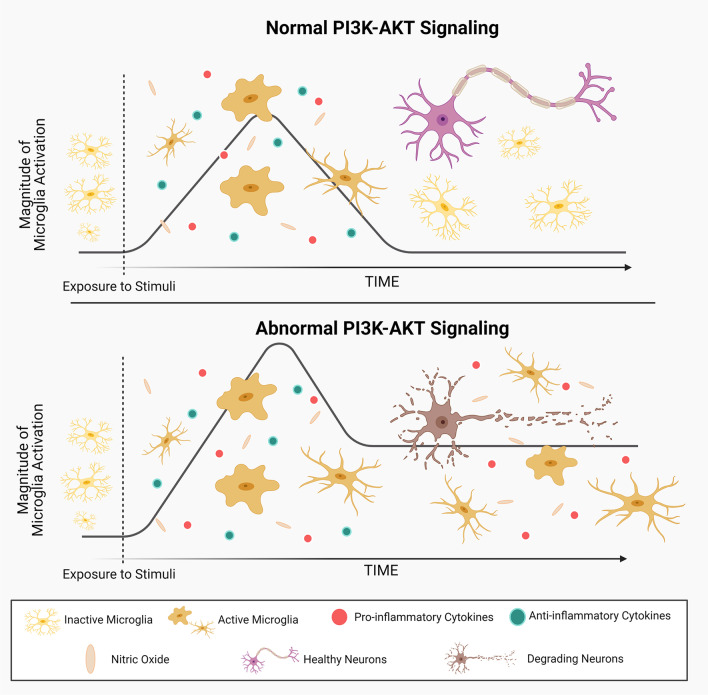
Microglia are integral mediators of innate immunity within the mammalian central nervous system. Typical microglial responses are transient, intending to restore homeostasis by orchestrating the removal of pathogens and debris and the regeneration of damaged neurons. However, prolonged and persistent microglial activation can drive chronic neuroinflammation and is associated with neurodegenerative disease. Recent evidence has revealed that abnormalities in microglial signaling pathways involving phosphatidylinositol 3-kinase (PI3K) and protein kinase B (AKT) may contribute to altered microglial activity and exacerbated neuroimmune responses. In this scoping review, the known and suspected roles of PI3K-AKT signaling in microglia, both during health and pathological states, will be examined, and the key microglial receptors that induce PI3K-AKT signaling in microglia will be described. Since aberrant signaling is correlated with neurodegenerative disease onset, the relationship between maladapted PI3K-AKT signaling and the development of neurodegenerative disease will also be explored. Finally, studies in which microglial PI3K-AKT signaling has been modulated will be highlighted, as this may prove to be a promising therapeutic approach for the future treatment of a range of neuroinflammatory conditions.

Keywords: AKT; Cell signaling; Glia; Innate immune system; Neurodegenerative diseases; PI3K.


Conflict of interest statement

The authors declare that there are no competing interests.

Figures

Fig. 1
Fig. 1
PI3K-AKT signaling pathway in microglia. PI3K is comprised of a p85 regulatory subunit that directs its location and a p110 catalytic subunit that phosphorylates phospholipids to mediate downstream signaling. In response to activation signals, PI3K is recruited to phosphotyrosine-containing motifs in cell surface receptors via the SH2 domain in p85, whereupon it acts on the membrane phospholipid, PI(4,5)P2, generating the second messenger PI(3,4,5)P3. PI(3,4,5)P3 recruits protein kinase B (AKT) to the membrane resulting in its activation, where it can phosphorylate numerous downstream substrates, leading to changes in gene transcription and biological responses. Upon receiving an inhibitory signal, the lipid phosphatase SHIP-1 is recruited to the membrane, where it converts PI(3,4,5)P3 into phosphatidylinositol 3,4 bisphosphate (PI(3,4)P2) to downmodulate signaling. PTEN is a direct antagonist of PI3K, converting PI(3,4,5)P3 to PI(4,5)P2. SFK Src family tyrosine kinase; Lyn Lck/yes-related novel tyrosine kinase; PH Pleckstrin homology domain
Fig. 2
Fig. 2
Key cell surface receptors in microglia that utilize PI3K-AKT signaling. CSF-1 or IL-34 binds to CSF-1R on microglia, triggering its dimerization, autophosphorylation and recruitment of PI3K. Endotoxin (LPS) binds to the TLR4 complex comprised of CD14 and MD-2, causing it to dimerize and induce recruitment of the adaptor MyD88, leading to its phosphorylation and the promotion of PI3K signaling. Neurotoxic plaques or other agonists bind to TREM2 and the co-associated membrane-bound adaptor protein DAP12 becomes phosphorylated on tyrosine-containing activation motifs (ITAMs), resulting in PI3K recruitment and activation. CX3CL1 binds to CX3CR1 and triggers the activation of heterotrimeric G proteins that transduce an intracellular signal via PI3K
Fig. 3
Fig. 3
Dysregulation of PI3K-AKT signaling causes abnormal microglial responses and promotes neuronal damage and chronic neuroinflammation. Resting microglia undergo activation upon encountering stimuli in their environment, triggering the release of nitric oxide and the coordinated expression of pro- and anti-inflammatory cytokines to clear the stimulus and promote recovery. Under normal conditions, PI3K-AKT signaling is terminated, the microglial response subsides and neurons are preserved. However, in certain settings, PI3K-AKT signaling is sustained, microglial responses persist perpetuating the release of pro-inflammatory cytokines into the brain microenvironment, which damages healthy neurons and contributes to progressive neurodegeneration

Similar articles

Cited by

References

    1. Janssen WJ, Henson PM. Cellular regulation of the inflammatory response. Toxicol Pathol. 2012;40(2):166–173. - PMC - PubMed
    1. Yang QQ, Zhou JW. Neuroinflammation in the central nervous system: symphony of glial cells. Glia. 2019;67(6):1017–1035. - PubMed
    1. Macháček T, Panská L, Dvořáková H, Horák P. Nitric oxide and cytokine production by glial cells exposed in vitro to neuropathogenic schistosome Trichobilharzia regenti. Parasit Vectors. 2016;9(1):579. - PMC - PubMed
    1. Lively S, Schlichter LC. Microglia responses to pro-inflammatory stimuli (LPS, IFNγ+TNFα) and reprogramming by resolving cytokines (IL-4, IL-10) Front Cell Neurosci. 2018 doi: 10.3389/fncel.2018.00215. - DOI - PMC - PubMed
    1. von Bartheld CS, Bahney J, Herculano-Houzel S. The search for true numbers of neurons and glial cells in the human brain: a review of 150 years of cell counting. J Comp Neurol. 2016;524(18):3865–3895. - PMC - PubMed

Substances

LinkOut - more resources