

Retina-attached slice recording reveals light-triggered tonic GABA signaling in suprachiasmatic nucleus
- PMID: 34838118
- PMCID: PMC8626980
- DOI: 10.1186/s13041-021-00881-9
Retina-attached slice recording reveals light-triggered tonic GABA signaling in suprachiasmatic nucleus
Abstract
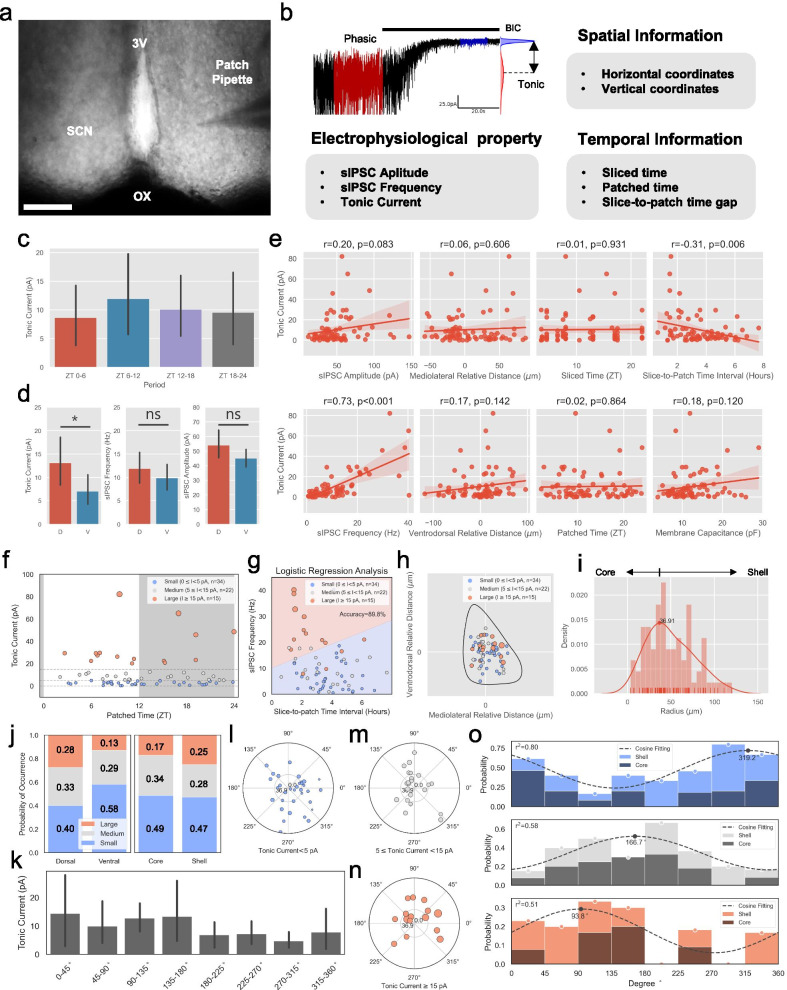
Light is a powerful external cue modulating the biological rhythm of internal clock neurons in the suprachiasmatic nucleus (SCN). GABA signaling in SCN is critically involved in this process. Both phasic and tonic modes of GABA signaling exist in SCN. Of the two modes, the tonic mode of GABA signaling has been implicated in light-mediated synchrony of SCN neurons. However, modulatory effects of external light on tonic GABA signalling are yet to be explored. Here, we systematically characterized electrophysiological properties of the clock neurons and determined the spatio-temporal profiles of tonic GABA current. Based on the whole-cell patch-clamp recordings from 76 SCN neurons, the cells with large tonic GABA current (>15 pA) were more frequently found in dorsal SCN. Moreover, tonic GABA current in SCN was highly correlated with the frequency of spontaneous inhibitory postsynaptic current (sIPSC), raising a possibility that tonic GABA current is due to spill-over from synaptic release. Interestingly, tonic GABA current was inversely correlated with slice-to-patch time interval, suggesting a critical role of retinal light exposure in intact brain for an induction of tonic GABA current in SCN. To test this possibility, we obtained meticulously prepared retina-attached SCN slices and successfully recorded tonic and phasic GABA signaling in SCN neurons. For the first time, we observed an early-onset, long-lasting tonic GABA current, followed by a slow-onset, short-lasting increase in the phasic GABA frequency, upon direct light-illumination of the attached retina. This result provides the first evidence that external light cue can directly trigger both tonic and phasic GABA signaling in SCN cell. In conclusion, we propose tonic GABA as the key mediator of external light in SCN.
Keywords: Light triggered tonic GABA; Retina-attached SCN; Spatiotemporal analysis; Whole-cell patch.
© 2021. The Author(s).
Conflict of interest statement
The authors declare that they have no competing interests.
Figures

Similar articles
-
Pre- and postsynaptic GABA(B) receptors modulate rapid neurotransmission from suprachiasmatic nucleus to parvocellular hypothalamic paraventricular nucleus neurons.Neuroscience. 2003;118(1):49-58. doi: 10.1016/s0306-4522(02)00906-5. Neuroscience. 2003. PMID: 12676136
-
Circadian modulation of GABA function in the rat suprachiasmatic nucleus: excitatory effects during the night phase.J Neurophysiol. 2002 Feb;87(2):834-44. doi: 10.1152/jn.00241.2001. J Neurophysiol. 2002. PMID: 11826050
-
Circadian rhythm in inhibitory synaptic transmission in the mouse suprachiasmatic nucleus.J Neurophysiol. 2004 Jul;92(1):311-9. doi: 10.1152/jn.01078.2003. Epub 2004 Feb 18. J Neurophysiol. 2004. PMID: 14973316 Free PMC article.
-
GABAergic mechanisms in the suprachiasmatic nucleus that influence circadian rhythm.J Neurochem. 2021 Apr;157(1):31-41. doi: 10.1111/jnc.15012. Epub 2020 Jul 3. J Neurochem. 2021. PMID: 32198942 Review.
-
The dynamics of GABA signaling: Revelations from the circadian pacemaker in the suprachiasmatic nucleus.Front Neuroendocrinol. 2017 Jan;44:35-82. doi: 10.1016/j.yfrne.2016.11.003. Epub 2016 Nov 25. Front Neuroendocrinol. 2017. PMID: 27894927 Free PMC article. Review.
Cited by
-
Immediate responses to ambient light in vivo reveal distinct subpopulations of suprachiasmatic VIP neurons.iScience. 2023 Sep 9;26(10):107865. doi: 10.1016/j.isci.2023.107865. eCollection 2023 Oct 20. iScience. 2023. PMID: 37766975 Free PMC article.
-
Evaluation of GABA Production by Alginate-Microencapsulated Fresh and Freeze-Dried Bacteria Enriched with Monosodium Glutamate during Storage in Chocolate Milk.Microorganisms. 2023 Oct 28;11(11):2648. doi: 10.3390/microorganisms11112648. Microorganisms. 2023. PMID: 38004660 Free PMC article.
-
GABA tone regulation and its cognitive functions in the brain.Nat Rev Neurosci. 2023 Sep;24(9):523-539. doi: 10.1038/s41583-023-00724-7. Epub 2023 Jul 26. Nat Rev Neurosci. 2023. PMID: 37495761 Review.
References
-
- Kim DY, Choi HJ, Kim JS, Kim YS, Jeong DU, Shin HC, Kim MJ, Han H-C, Hong SK, Kim YI. Voltage-gated calcium channels play crucial roles in the glutamate-induced phase shifts of the rat suprachiasmatic circadian clock. Eur J Neurosci. 2005;21(5):1215–1222. doi: 10.1111/j.1460-9568.2005.03950.x. - DOI - PubMed
Publication types
MeSH terms
Substances
LinkOut - more resources
Full Text Sources
