. 2022 Jan:197:105212.
doi: 10.1016/j.antiviral.2021.105212. Epub 2021 Nov 24.

Hydroxychloroquine and azithromycin used alone or combined are not effective against SARS-CoV-2 ex vivo and in a hamster model

Affiliations

Hydroxychloroquine and azithromycin used alone or combined are not effective against SARS-CoV-2 ex vivo and in a hamster model

Maxime Cochin et al. Antiviral Res. 2022 Jan.

Abstract

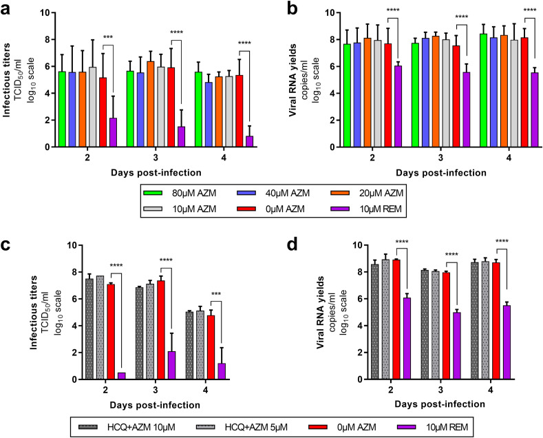
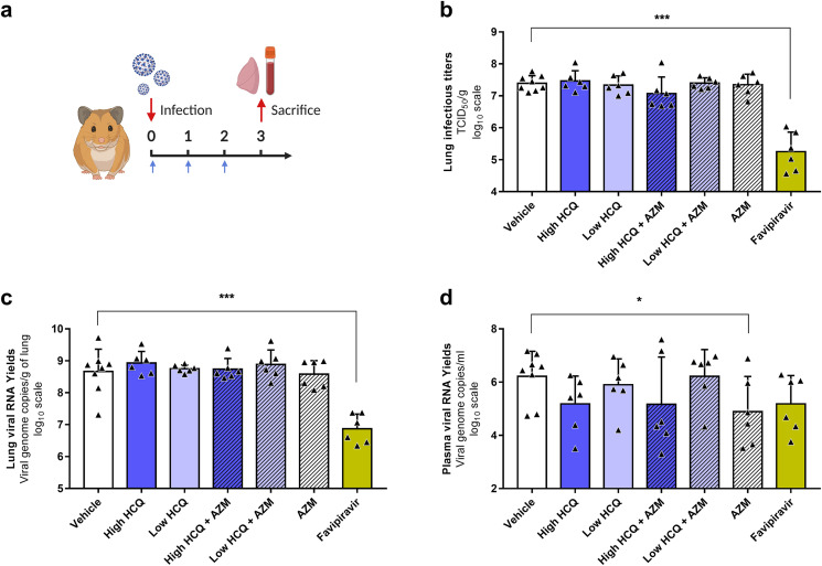
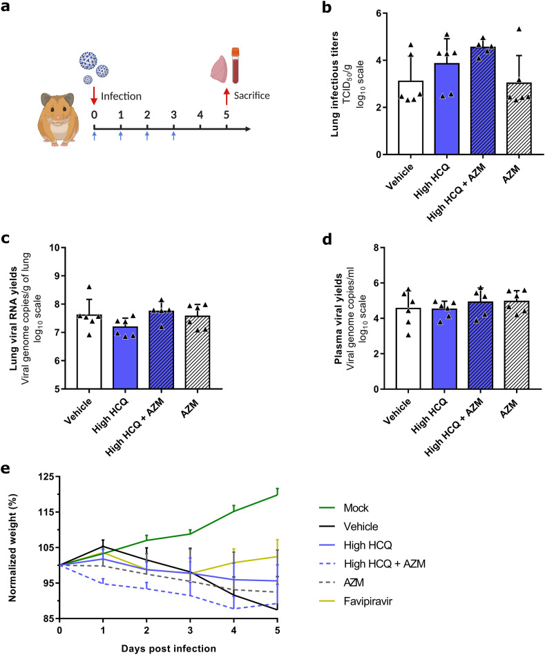
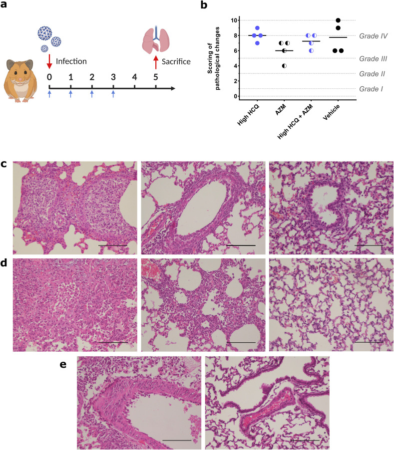
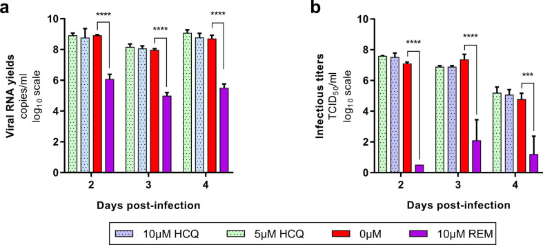
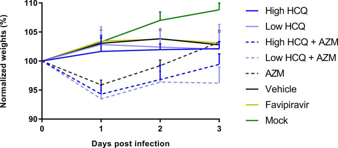
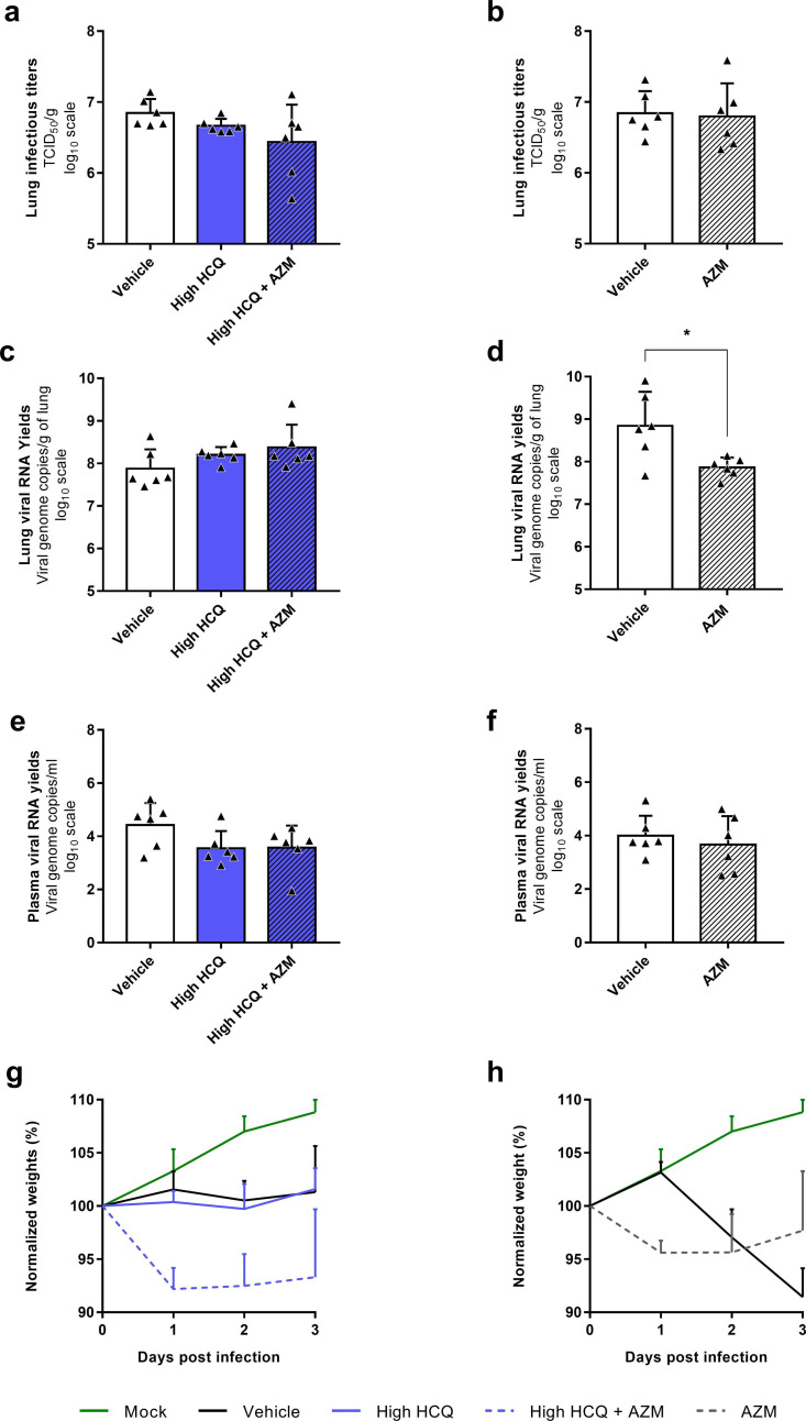
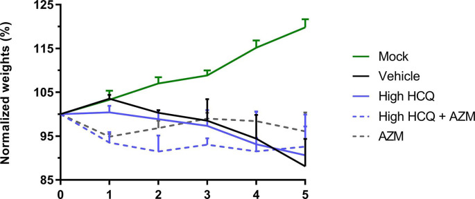
Drug repositioning has been used extensively since the beginning of the COVID-19 pandemic in an attempt to identify antiviral molecules for use in human therapeutics. Hydroxychloroquine and azithromycin have shown inhibitory activity against SARS-CoV-2 replication in different cell lines. Based on such in vitro data and despite the weakness of preclinical assessment, many clinical trials were set up using these molecules. In the present study, we show that hydroxychloroquine and azithromycin alone or combined does not block SARS-CoV-2 replication in human bronchial airway epithelia. When tested in a Syrian hamster model, hydroxychloroquine and azithromycin administrated alone or combined displayed no significant effect on viral replication, clinical course of the disease and lung impairments, even at high doses. Hydroxychloroquine quantification in lung tissues confirmed strong exposure to the drug, above in vitro inhibitory concentrations. Overall, this study does not support the use of hydroxychloroquine and azithromycin as antiviral drugs for the treatment of SARS-CoV-2 infections.

Keywords: Antivirals; Azithromycin; COVID-19; Coronavirus; Ex vivo; Human airway epithelium; Hydroxychloroquine; In vivo; SARS-CoV-2; Syrian hamster.


Conflict of interest statement

The authors declare that they have no known competing financial interests or personal relationships that could have appeared to influence the work reported in this paper.

Figures

Fig. 1
Fig. 1
Antiviral activity of AZM and AZM-HCQ combination in a bronchial human airway epithelium (HAE). Kinetics of virus excretion at the apical side of the epithelium in presence of AZM (a,b) and HCQ-AZM combination (c,d). Kinetics were measured using a TCID50 assay (a,c) and a RT-qPCR assay (b,d). For panels a and b, data represent mean ± SD of two independent experiments each performed in duplicate (n = 4). For panels c and d, data represent mean ± SD of on experiment performed in triplicate (details in Supplemental Table 2). **** and *** symbols indicate that infectious titers or RNA yields are significantly smaller than those for untreated (0 μM AZM) epithelia with a p-value inferior to 0.0001 or ranging between 0.0001 and 0.001, respectively (Multiple comparison t-test) (details in Supplemental Table 3).
Fig. 2
Fig. 2
Antiviral activity of HCQ and AZM alone or combined in a Syrian hamster model at 3 dpi. (a) Experimental timeline (realized on biorender.com). Groups of 6 or 8 hamsters were intranasally infected with 1 × 104 TCID50 of SARS-CoV-2, treated from 0 to 2 dpi (blue arrows) and sacrificed at 3 dpi. (b) Lung infectious titers measured using a TCID50 assay. (c–d) Lung viral RNA yields (c) and plasma viral loads (d) measured using a RT-qPCR assay. Data represent mean ± SD of individual data of hamsters (n = 6 to 8; details in Supplemental Table 4). *** and * symbols indicate that lung infectious titers, lung viral RNA yields or plasma viral loads are significantly smaller than those for the untreated group (vehicle) with a p-value ranging between 0.0001-0.001 and 0.01–0.05, respectively (Unpaired and Welch's t tests). Clinical follow-up of this experiment is presented in Supplemental Fig. 3.
Fig. 3
Fig. 3
Antiviral activity and clinical impact of HCQ and AZM alone or combined in a Syrian hamster model at 5 dpi. (a) Experimental timeline (realized on biorender.com). Groups of 6 hamsters were intranasally infected with 1 × 104 TCID50 of SARS-CoV-2, treated from 0 to 3 dpi (blue arrows) and sacrificed at 5 dpi. (b) Lung infectious titers measured using a TCID50 assay. (c–d) Lung viral RNA yields (c) and plasma viral loads (d) measured using a RT-qPCR assay. (e) Clinical follow-up. Animal weights are expressed as normalized weights (i.e. % of initial weight). Data represent mean ± SD of individual data of hamsters (n = 5 to 6; details in Supplemental Table 4)
Fig. 4
Fig. 4
Impact of HCQ and AZM alone or combined on lung histological impairments in a Syrian hamster model at 5 dpi. (a) Experimental timeline (realized on biorender.com). Groups of 4 hamsters were intranasally infected with 1 × 104 TCID50 of SARS-CoV-2, treated from 0 to 3 dpi and sacrificed at 5 dpi (blue arrows). (b) Scores of histopathological changes measured following criteria presented in Supplemental Table 1, as previously described (Driouich et al., 2021). Dashes represent mean scores for each group (n = 4; details in Supplemental Table 5). (c) Representative images of bronchial inflammation (scale bar: 100 μ). From left to right, pictures represent marked neutrophilic bronchitis (high HCQ + AZM animal), mild peribronchial leucocytic infiltration (high HCQ + AZM animal) and no inflammation (mock-infected animal). (d) Representative images of alveolar inflammation (scale bar: 100 μ). From left to right, pictures represent severe interstitial pneumonia (high HCQ animal), marked interstitial changes (high HCQ + AZM animal) and no inflammation (mock-infected animal). (e) Representative images of vessel changes (leukocyte accumulation within vascular walls). From left to right, pictures represent presence (high HCQ animal) and absence of lesions (mock-infected animal) (scale bars: 100 μ). Clinical follow-up is represented in Supplemental Fig. 5.
None
None
None
None
None

References

    1. Andreani J., Le Bideau M., Duflot I., Jardot P., Rolland C., Boxberger M., Wurtz N., Rolain J.M., Colson P., La Scola B., Raoult D. In vitro testing of combined hydroxychloroquine and azithromycin on SARS-CoV-2 shows synergistic effect. Microb. Pathog. 2020;145:104228. - PMC - PubMed
    1. Barnard D.L., Day C.W., Bailey K., Heiner M., Montgomery R., Lauridsen L., Chan P.K., Sidwell R.W. Evaluation of immunomodulators, interferons and known in vitro SARS-coV inhibitors for inhibition of SARS-coV replication in BALB/c mice. Antivir. Chem. Chemother. 2006;17:275–284. - PubMed
    1. Baronas E.T., Lee J.-W., Alden C., Hsieh F.Y. Biomarkers to monitor drug-induced phospholipidosis. Toxicol. Appl. Pharmacol. 2007;218:72–78. - PubMed
    1. Beigelman A., Mikols C.L., Gunsten S.P., Cannon C.L., Brody S.L., Walter M.J. Azithromycin attenuates airway inflammation in a mouse model of viral bronchiolitis. Respir. Res. 2010;11:90. - PMC - PubMed
    1. Browning D.J. Springer New York; New York, NY: 2014. Pharmacology of Chloroquine and Hydroxychloroquine, Hydroxychloroquine and Chloroquine Retinopathy; pp. 35–63.

MeSH terms