. 2022 Feb:142:106946.
doi: 10.1016/j.vph.2021.106946. Epub 2021 Nov 25.

Mitochondrial DNA and TLR9 activation contribute to SARS-CoV-2-induced endothelial cell damage

Affiliations

Mitochondrial DNA and TLR9 activation contribute to SARS-CoV-2-induced endothelial cell damage

Tiago J Costa et al. Vascul Pharmacol. 2022 Feb.

Abstract

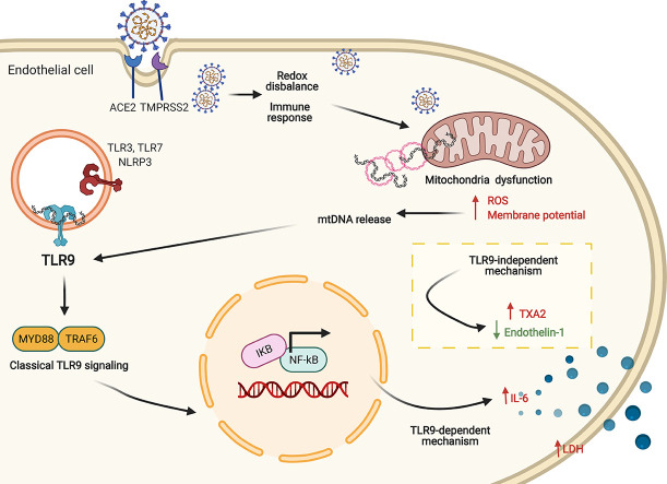
Background and purpose: Mitochondria play a central role in the host response to viral infection and immunity, being key to antiviral signaling and exacerbating inflammatory processes. Mitochondria and Toll-like receptor (TLR) have been suggested as potential targets in SARS-CoV-2 infection. However, the involvement of TLR9 in SARS-Cov-2-induced endothelial dysfunction and potential contribution to cardiovascular complications in COVID-19 have not been demonstrated. This study determined whether infection of endothelial cells by SARS-CoV-2 affects mitochondrial function and induces mitochondrial DNA (mtDNA) release. We also questioned whether TLR9 signaling mediates the inflammatory responses induced by SARS-CoV-2 in endothelial cells.

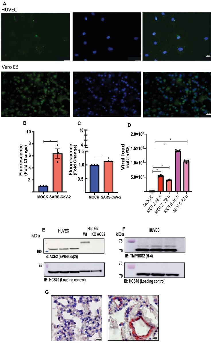
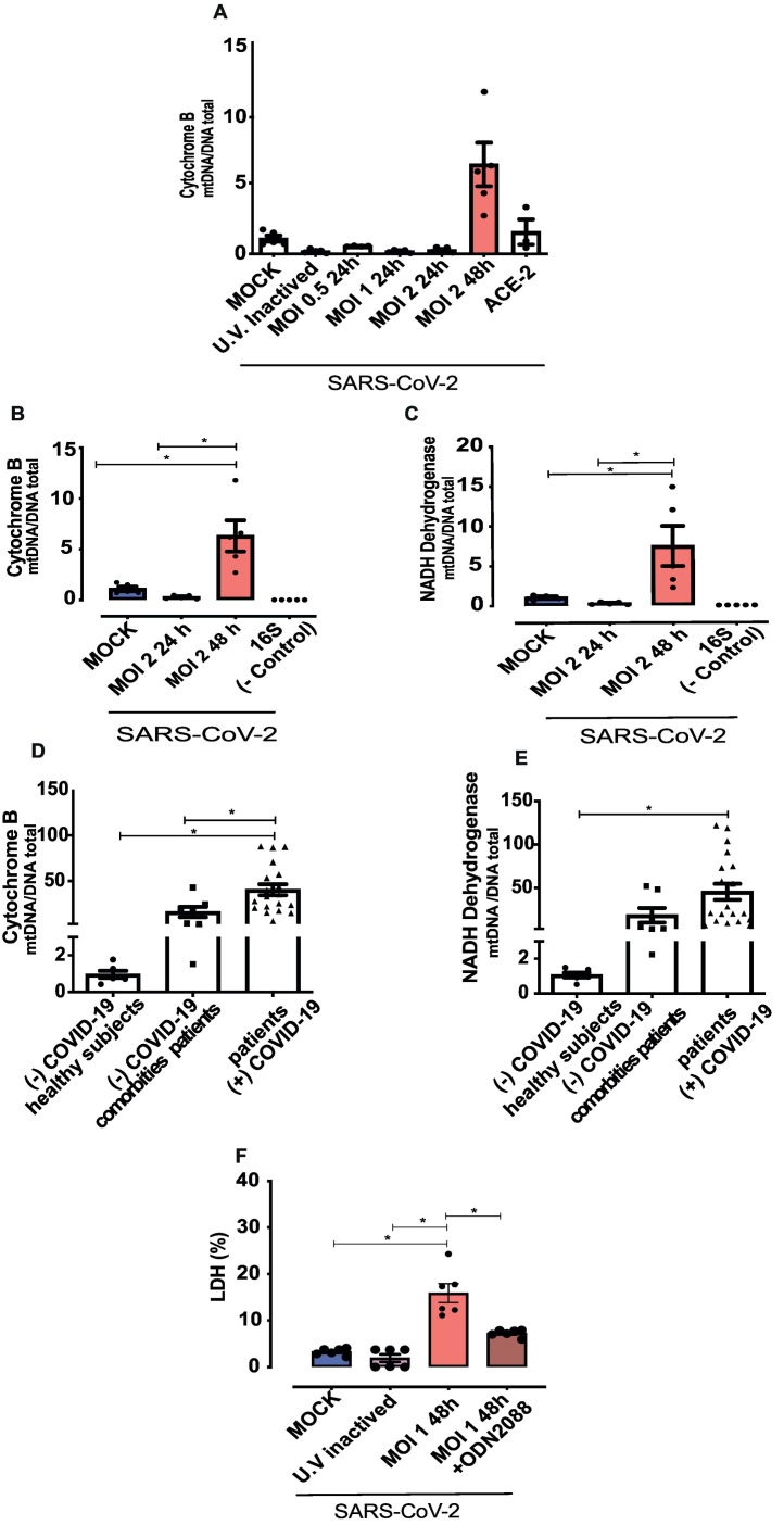
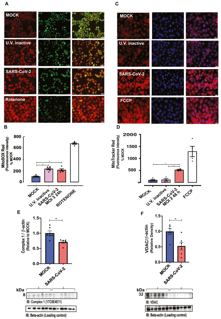
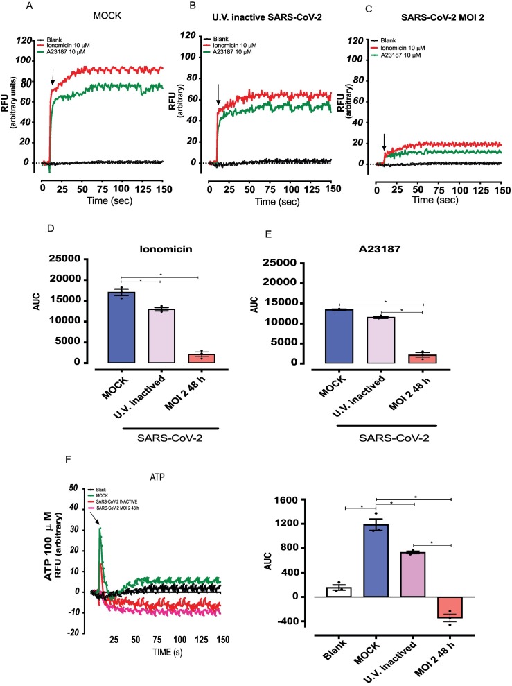
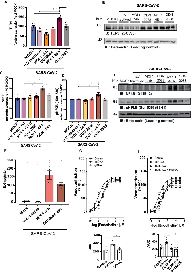
Experimental approach: Human umbilical vein endothelial cells (HUVECs) were infected by SARS-CoV-2 and immunofluorescence was used to confirm the infection. Mitochondrial function was analyzed by specific probes and mtDNA levels by real-time polymerase chain reaction (RT-PCR). Inflammatory markers were measured by ELISA, protein expression by western blot, intracellular calcium (Ca2+) by FLUOR-4, and vascular reactivity with a myography.

Key results: SARS-CoV-2 infected HUVECs, which express ACE2 and TMPRSS2 proteins, and promoted mitochondrial dysfunction, i.e. it increased mitochondria-derived superoxide anion, mitochondrial membrane potential, and mtDNA release, leading to activation of TLR9 and NF-kB, and release of cytokines. SARS-CoV-2 also decreased nitric oxide synthase (eNOS) expression and inhibited Ca2+ responses in endothelial cells. TLR9 blockade reduced SARS-CoV-2-induced IL-6 release and prevented decreased eNOS expression. mtDNA increased vascular reactivity to endothelin-1 (ET-1) in arteries from wild type, but not TLR9 knockout mice. These events were recapitulated in serum samples from COVID-19 patients, that exhibited increased levels of mtDNA compared to sex- and age-matched healthy subjects and patients with comorbidities.

Conclusion and applications: SARS-CoV-2 infection impairs mitochondrial function and activates TLR9 signaling in endothelial cells. TLR9 triggers inflammatory responses that lead to endothelial cell dysfunction, potentially contributing to the severity of symptoms in COVID-19. Targeting mitochondrial metabolic pathways may help to define novel therapeutic strategies for COVID-19.

Keywords: Endothelial dysfunction; Mitochondria; SARS-CoV-2; Toll like receptor 9.


Conflict of interest statement

The authors declare no conflict of interest.

Figures

Unlabelled Image
Graphical abstract
Fig. 1
Fig. 1
Endothelial cells are infected by SARS-CoV-2. (A) Immunofluorescence in HUVECs (top panels) and Vero-E6 cells (positive control, bottom panels) infected with SARS-CoV-2 (GFP signals were detected at 48 h post-infection). (B and C) fluorescence intensity (FI) in the region of interest (ROI) was determined using the Image J software in HUVECs and Vero-E6 cells, respectively. (D) Viral load in endothelial cells 48 and 72 h post-infection (MOI 2 and 5). (E) Representative immunoblot image of ACE2 protein in HUVECs and Hep G2 cells, wildtype and submitted to ACE2 deletion by CRISPR-Cas9. (F) Representative immunoblot image of TMPRSS2 protein in HUVECs. HCS70 was used as the loading control. (G) Immunochemistry for SARS-CoV-2 in lung samples of COVID-19 positive (right) and COVID-19 negative (left) patients (Scale bars, 10 μm.). The results are expressed as the mean ± SEM. Statistical significance was determined by unpaired test t-test (n = 3) or one-way ANOVA multiple comparations using the Prism GraphPad 8.0 software. Statistically significant differences were considered when p < 0.05. MOCK, MOI, multiplicity of infection.
Fig. 2
Fig. 2
SARS-CoV-2 induces endothelial cell damage and mitochondrial DNA release. (A) Time and MOI-dependent infection of endothelial cells bySARS-CoV-2. (B and C), cytochrome B and NADH dehydrogenase expression in the supernatant of SARS-CoV-2-infected HUVECs. (D and E) Cytochrome B and NADH dehydrogenase analysis in the serum of positive and negative COVID-19 patients. (F) Lactate dehydrogenase (LDH) assay to identify cell damage. Results are expressed as % of control. NADH dehydrogenase and cytochrome B values are expressed as mtDNA/total DNA). The results are expressed as the mean ± SEM. Statistical significance was determined by one-way ANOVA followed by the multiple comparations Tukey post-hoc test (cells n = 3–5; patients n = 7–19), using the Prism GraphPad 8.0 software. Statistically significant differences were considered when p < 0.05.
Fig. 3
Fig. 3
SARS-CoV-2 increases mitochondrial dysfunction. Fluorescence intensity depicting (A and B) mitochondrial ROS (MitoSOX Red®) and (C and D) mitochondrial potential (MitoTracker® Red CMXRos, FlexStation-3) in control (MOCK and UV-inactivated SARS-CoV-2-exposed HUVECs) and SARS-CoV-2-infected (MOI 2–24 or 48 h) HUVECs. Rotenone and FCCP were used as positive controls. Acridine orange (green) and DAPI (blue) were utilized for the nucleus staining. Immunoblot representative imagens (bottom) and densitometric analysis (top) of protein levels of (E) mitochondrial complex I and (F) VDAC (voltage-dependent ion channel) normalized by β-actin in MOCK and SARS-CoV-2-infected HUVECs. The results are expressed as the mean ± SEM. Statistical significance was determined by one-way ANOVA multiple comparations with Tukey post-hoc test or t-test when appropriated (n = 5–6), using the Prism GraphPad 8.0 software. Statistically significant differences were considered when p < 0.05. (For interpretation of the references to colour in this figure legend, the reader is referred to the web version of this article.)
Fig. 4
Fig. 4
SARS-CoV-2 decreases calcium responses in endothelial cells. Calcium mobilization in response to ionomycin (10−5 M) and A23187 (10−6 M) in HUVECs cells in (A) free MOCK medium, (B) exposed to UV-inactivated SARS-CoV-2, (C) SARS-CoV-2 (MOI 2) for 48 h. (D) and (E) represent the AUC. (F and G) Calcium mobilization in response to ATP. The results are expressed as the mean ± SEM. Statistical significance was determined by one-way ANOVA multiple comparations with Tukey post-hoc test (n = 3), using the Prism GraphPad 8.0 software. Statistically significant differences were considered when p < 0.05.
Fig. 5
Fig. 5
SARS-CoV-2 increases protein levels of TLR9 and NF-kB signaling. Representative immunoblot images and densitometric analysis of protein levels of (A and B) TLR9, (C, D and E) total and phosphorylated (Serine536) NF-kB in HUVECs infected with SARS-CoV-2 for 24 and 48 h treated with vehicle or ODN 2088, TLR9 antagonist. Expression of the protein of interest was normalized by β-actin expression. Results are expressed as a percentage of MOCK control and represent the mean ± SEM from 4 experiments. The results are expressed as the mean ± SEM. Statistical significance was determined by one-way ANOVA multiple comparations with Tukey post-hoc test (n = 4), using the Prism GraphPad 8.0 software. Statistically significant differences were considered when p < 0.05. (F) IL-6 determined by Elisa assay in the supernatant of HUVECs infected by SARS-CoV-2; values represent pg/mL. Cumulative concentration-response curves to endothelin-1 in resistance mesenteric arteries of C57BL/J6 (G) and TLR9KO (H) mice, in the presence of mtDNA (mitochondrial DNA) or gDNA (genomic DNA). Results are expressed as a percentage of KCl-induced vasoconstriction and represent the mean ± SEM from 3 to 4 different experiments. Statistical significance was determined by one-way ANOVA multiple comparations with Tukey post-hoc test (n = 3–6), using the Prism GraphPad 8.0 software. Statistically significant differences were considered when p < 0.05.

References

    1. CSSE-JHU, C. F. S. S. A. E. A. J. H. U COVID-19 Dashboard n.d. 2020. https://coronavirus.jhu.edu/map.html
    1. Menter T., et al. Postmortem examination of COVID-19 patients reveals diffuse alveolar damage with severe capillary congestion and variegated findings in lungs and other organs suggesting vascular dysfunction. Histopathology. Aug 2020;77(2):198–209. https://www.ncbi.nlm.nih.gov/pubmed/32364264 ISSN 1365–2559. Disponível em. - PMC - PubMed
    1. Li S., et al. SARS-CoV-2 triggers inflammatory responses and cell death through caspase-8 activation. Signal Transduct Target Ther. 2020;5(1):235. https://www.ncbi.nlm.nih.gov/pubmed/33037188 ISSN 2059–3635. Disponível em. - PMC - PubMed
    1. Dorward D.A., et al. Tissue-Specific Immunopathology in Fatal COVID-19. Am. J. Respir. Crit. Care Med. 2021;203(2):192–201. https://www.ncbi.nlm.nih.gov/pubmed/33217246 ISSN 1535–4970. Disponível em. - PMC - PubMed
    1. Rodrigues T.S., et al. Inflammasomes are activated in response to SARS-CoV-2 infection and are associated with COVID-19 severity in patients. J. Exp. Med. 2021;218(3) https://www.ncbi.nlm.nih.gov/pubmed/33231615 ISSN 1540–9538. Disponível em. - PMC - PubMed

Publication types