

Postmortem fluorescence angiography of the explanted human heart
- PMID: 34839382
- PMCID: PMC8813811
- DOI: 10.1007/s00414-021-02730-9
Postmortem fluorescence angiography of the explanted human heart
Abstract
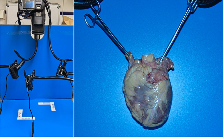
Within the scope of this technical report, the feasibility of indocyanine green (ICG) as a fluorescent agent for postmortem angiography of the heart is tested. The study included 4 deceased persons with no respective medical history of heart diseases. The basic patterns of findings in ICG fluorescence angiography associated with healthy hearts are presented. The method can easily be integrated into a workflow without restricting the macroscopic or histologic diagnostics. This paper represents the fundamental technical and analytical basis for upcoming studies concerning the possibilities and limitations of fluorescence angiography in the diagnosis of heart pathology.
Keywords: Angiography; Fluorescence; Heart; ICG; Indocyanine green; Postmortem.
© 2021. The Author(s).
Conflict of interest statement
The authors declare no competing interests.
Figures

References
-
- Riße M, Weiler G. Coronary muscle bridge and its relations to local coronary sclerosis, regional myocardial ischemia and coronary spasm. A morphometric study. (article in German) Z Kardiol. 1985;74:700–705. - PubMed
-
- Weiler G. Quantitative morphology of the coronary arteries in acute coronary death. (article in German) Fortschr Med. 1980;98:580–583. - PubMed
-
- Campbell JS. Stereroscopic radiography of the coronary circulation. The Lancet. 1928;212:168–169. doi: 10.1016/S0140-6736(00)83656-9. - DOI
MeSH terms
Substances
LinkOut - more resources
Full Text Sources
