Review
. 2022 Jul:127:37-45.
doi: 10.1016/j.semcdb.2021.11.010. Epub 2021 Nov 25.

Mesoderm induction and patterning: Insights from neuromesodermal progenitors

Affiliations
Review

Mesoderm induction and patterning: Insights from neuromesodermal progenitors

Benjamin L Martin. Semin Cell Dev Biol. 2022 Jul.

Abstract

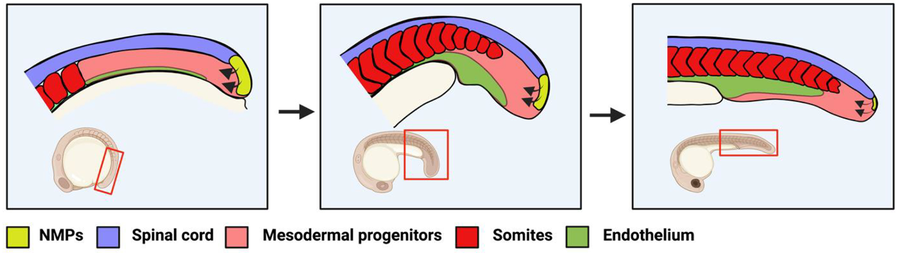
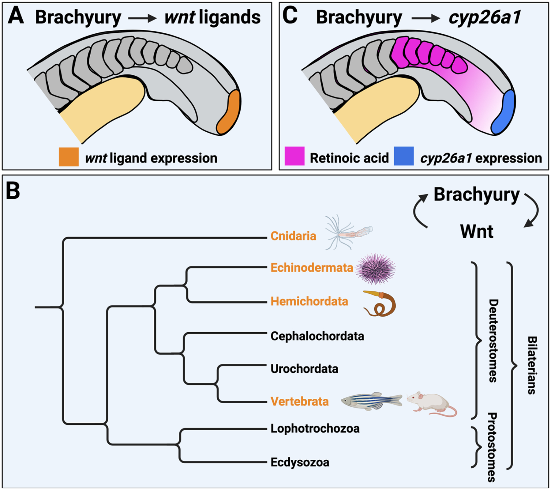
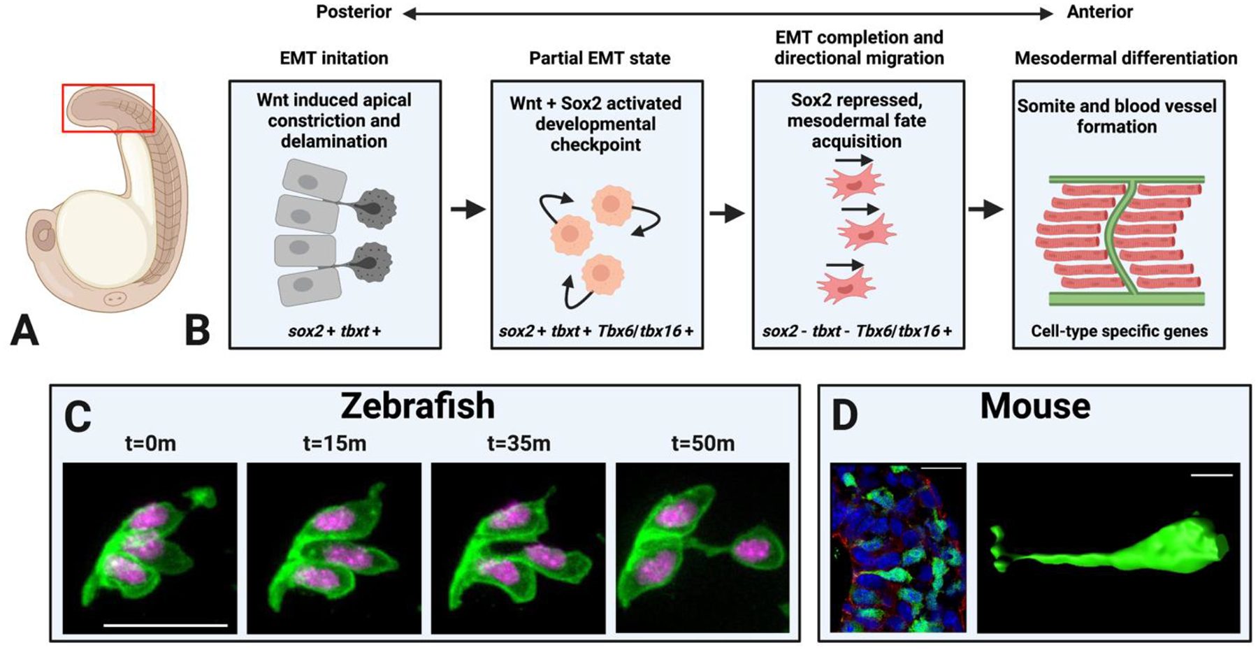
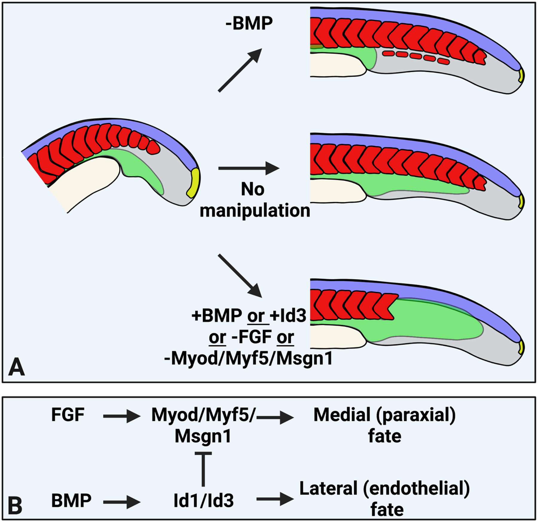
The discovery of mesoderm inducing signals helped usher in the era of molecular developmental biology, and today the mechanisms of mesoderm induction and patterning are still intensely studied. Mesoderm induction begins during gastrulation, but recent evidence in vertebrates shows that this process continues after gastrulation in a group of posteriorly localized cells called neuromesodermal progenitors (NMPs). NMPs reside within the post-gastrulation embryonic structure called the tailbud, where they make a lineage decision between ectoderm (spinal cord) and mesoderm. The majority of NMP-derived mesoderm generates somites, but also contributes to lateral mesoderm fates such as endothelium. The discovery of NMPs provides a new paradigm in which to study vertebrate mesoderm induction. This review will discuss mechanisms of mesoderm induction within NMPs, and how they have informed our understanding of mesoderm induction more broadly within vertebrates as well as animal species outside of the vertebrate lineage. Special focus will be given to the signaling networks underlying NMP-derived mesoderm induction and patterning, as well as emerging work on the significance of partial epithelial-mesenchymal states in coordinating cell fate and morphogenesis.

Keywords: BMP; Brachyury; EMT; Epithelial-mesenchymal transition; FGF; Mesoderm induction; Mesoderm patterning; Neuromesodermal progenitors; Wnt.


Figures

Figure 1 –
Figure 1 –. Neuromesodermal progenitors generate spinal cord and mesoderm after gastrulation.
Vertebrate embryos (here is shown a zebrafish embryo) contain NMPs (yellow) in the tailbud which generate spinal cord (purple) and mesodermal progenitors (pink). The mesoderm will differentiate into somites (red) and vascular endothelium (green). The process of mesoderm induction from NMPs occurs continuously (arrows) over the course of axial extension until the NMPs are depleted. The red boxed region of the inset embryo represents the area depicted in the schematics.
Figure 2 –
Figure 2 –. Conserved direct Brachyury targets involved in NMP maintenance and NMP-derived mesoderm induction.
(A) Brachyury directly activates the expression of canonical Wnt ligands in the NMP region (orange). Wnt signaling in turn activates brachyury expression. (B) A simplified animal phylogeny illustrating the groups in which there is experimental evidence for a Brachyury/Wnt autoregulatory loop (orange). The specific animals in which this has been observed are shown as icons next to the phylogenetic tree. (C) Brachyury target gene cyp26a1 is expressed in the NMPs (blue). Cyp26a1 degrades retinoic acid to protect NMPs from the nearby posterior somite retinoic acid source (magenta).
Figure 3 –
Figure 3 –. Insights into the mesodermal epithelial to mesenchymal transition.
(A) A zebrafish embryo depicting (red box) the region of interest shown in (B). (B) The stages of the NMP-derived mesodermal EMT are depicted as schematics with events occurring beginning in the posterior wall of the tailbud (left) and progressing towards the anterior (right). The marker genes expressed at each step are indicated under each schematic. (C) Micrographs from a time-lapse of an individual cell undergoing EMT during zebrafish NMP-derived mesoderm induction, adapted from [16] with permission. Scale bar = 20μm. (D) Micrograph (left) and surface rendering (right) of an individual cell undergoing EMT during gastrula stage mesoderm induction in a mouse embryo, adapted from [94] with permission. Scale bar in left image = 25μm, in right image = 10μm). Both the mouse and zebrafish cells exhibit apical constriction and eventual delamination from the epithelium.
Figure 4 –
Figure 4 –. Patterning of mesoderm by modulation of bHLH transcription factor activity.
(A) Experimental manipulations that change the patterning of NMP-derived mesoderm in vivo, resulting in loss of endothelium and formation of ectopic somite tissue (top), or loss of somites and expansion of endothelium (bottom). (B) A model depicting the molecular mechanism of patterning downstream of BMP and FGF signaling. FGF signaling activates bHLH transcription factor expression, while BMP signaling activates the Id genes which inhibit bHLH proteins. This mechanism has been shown in zebrafish and in cultured mouse NMP-derived mesoderm.

References

    1. Kimelman D, Martin BL. Anterior-posterior patterning in early development: three strategies. Wiley Interdiscip Rev Dev Biol 1(2) (2012) 253–66. - PMC - PubMed
    1. Tzouanacou E, Wegener A, Wymeersch FJ, Wilson V, Nicolas J-F. Redefining the Progression of Lineage Segregations during Mammalian Embryogenesis by Clonal Analysis. Developmental cell 17(3) (2009) 365–376. - PubMed
    1. Martin BL, Kimelman D. Canonical Wnt signaling dynamically controls multiple stem cell fate decisions during vertebrate body formation. Dev Cell 22(1) (2012) 223–32. - PMC - PubMed
    1. Kinney BA, Al Anber A, Row RH, Tseng YJ, Weidmann MD, Knaut H, Martin BL. Sox2 and Canonical Wnt Signaling Interact to Activate a Developmental Checkpoint Coordinating Morphogenesis with Mesoderm Fate Acquisition. Cell Rep 33(4) (2020) 108311. - PMC - PubMed
    1. Guillot C, Djeffal Y, Michaut A, Rabe B, Pourquie O. Dynamics of primitive streak regression controls the fate of neuromesodermal progenitors in the chicken embryo. Elife 10 (2021). - PMC - PubMed

Publication types

LinkOut - more resources