

Feasibility of Radiomics to Differentiate Coronavirus Disease 2019 (COVID-19) from H1N1 Influenza Pneumonia on Chest Computed Tomography: A Proof of Concept
- PMID: 34840382
- PMCID: PMC8611216
- DOI: 10.30476/ijms.2021.88036.1858
Feasibility of Radiomics to Differentiate Coronavirus Disease 2019 (COVID-19) from H1N1 Influenza Pneumonia on Chest Computed Tomography: A Proof of Concept
Abstract
Background: Chest computed tomography (CT) plays an essential role in diagnosing coronavirus disease 2019 (COVID-19). However, CT findings are often nonspecific among different viral pneumonia conditions. The differentiation between COVID-19 and influenza can be challenging when seasonal influenza concurs with the COVID-19 pandemic. This study was conducted to test the ability of radiomics-artificial intelligence (AI) to perform this task.
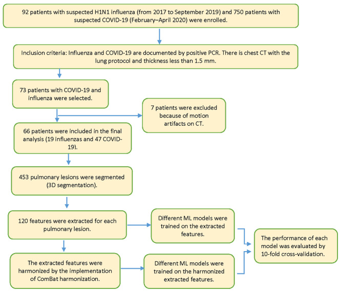
Methods: In this retrospective study, chest CT images from 47 patients with COVID-19 (after February 2020) and 19 patients with H1N1 influenza (before September 2019) pneumonia were collected from three hospitals affiliated with Arak University of Medical Sciences, Arak, Iran. All pulmonary lesions were segmented on CT images. Multiple radiomics features were extracted from the lesions and used to develop support-vector machine (SVM), k-nearest neighbor (k-NN), decision tree, neural network, adaptive boosting (AdaBoost), and random forest.
Results: The patients with COVID-19 and H1N1 influenza were not significantly different in age and sex (P=0.13 and 0.99, respectively). Nonetheless, the average time between initial symptoms/hospitalization and chest CT was shorter in the patients with COVID-19 (P=0.001 and 0.01, respectively). After the implementation of the inclusion and exclusion criteria, 453 pulmonary lesions were included in this study. On the harmonized features, random forest yielded the highest performance (area under the curve=0.97, sensitivity=89%, precision=90%, F1 score=89%, and classification accuracy=89%).
Conclusion: In our preliminary study, radiomics feature extraction, conjoined with AI, especially random forest and neural network, appeared to yield very promising results in the differentiation between COVID-19 and H1N1 influenza on chest CT.
Keywords: Artificial intelligence; COVID-19; Influenza, Human; Tomography.
Copyright: © Iranian Journal of Medical Sciences.
Figures
Similar articles
-
CT-based radiomics combined with signs: a valuable tool to help radiologist discriminate COVID-19 and influenza pneumonia.BMC Med Imaging. 2021 Feb 17;21(1):31. doi: 10.1186/s12880-021-00564-w. BMC Med Imaging. 2021. PMID: 33596844 Free PMC article.
-
A Comparison of Clinical and Chest CT Findings in Patients With Influenza A (H1N1) Virus Infection and Coronavirus Disease (COVID-19).AJR Am J Roentgenol. 2020 Nov;215(5):1065-1071. doi: 10.2214/AJR.20.23214. Epub 2020 May 26. AJR Am J Roentgenol. 2020. PMID: 32452731
-
CT Manifestations of Coronavirus Disease (COVID-19) Pneumonia and Influenza Virus Pneumonia: A Comparative Study.AJR Am J Roentgenol. 2021 Jan;216(1):71-79. doi: 10.2214/AJR.20.23304. Epub 2020 Jul 9. AJR Am J Roentgenol. 2021. PMID: 32755175
-
Chest Computed Tomography Findings in COVID-19 and Influenza: A Narrative Review.Biomed Res Int. 2020 Jun 5;2020:6928368. doi: 10.1155/2020/6928368. eCollection 2020. Biomed Res Int. 2020. PMID: 32596354 Free PMC article. Review.
-
Artificial intelligence model on chest imaging to diagnose COVID-19 and other pneumonias: A systematic review and meta-analysis.Eur J Radiol Open. 2022;9:100438. doi: 10.1016/j.ejro.2022.100438. Epub 2022 Aug 18. Eur J Radiol Open. 2022. PMID: 35996746 Free PMC article. Review.
Cited by
-
Quantitative Evaluation of COVID-19 Pneumonia CT Using AI Analysis-Feasibility and Differentiation from Other Common Pneumonia Forms.Diagnostics (Basel). 2023 Jun 20;13(12):2129. doi: 10.3390/diagnostics13122129. Diagnostics (Basel). 2023. PMID: 37371024 Free PMC article.
-
Development and Validation of a Radiomics Nomogram Using Computed Tomography for Differentiating Immune Checkpoint Inhibitor-Related Pneumonitis From Radiation Pneumonitis for Patients With Non-Small Cell Lung Cancer.Front Immunol. 2022 Apr 26;13:870842. doi: 10.3389/fimmu.2022.870842. eCollection 2022. Front Immunol. 2022. PMID: 35558076 Free PMC article.
-
Artificial intelligence-driven assessment of radiological images for COVID-19.Comput Biol Med. 2021 Sep;136:104665. doi: 10.1016/j.compbiomed.2021.104665. Epub 2021 Jul 21. Comput Biol Med. 2021. PMID: 34343890 Free PMC article. Review.
References
-
- Lake MA. What we know so far: COVID-19 current clinical knowledge and research. Clin Med (Lond) 2020;20:124–7. doi: 10.7861/clinmed.2019-coron. [ PMC Free Article ] - DOI - PMC - PubMed
-
- Johns Hopkins University [Interent] COVID-19 Dashboard by the Center for Systems Science and Engineering (CSSE). [Cited 8 September 2020] Available from: https://coronavirus.jhu.edu/map.html .
-
- Long C, Xu H, Shen Q, Zhang X, Fan B, Wang C, et al. Diagnosis of the Coronavirus disease (COVID-19): rRT-PCR or CT? Eur J Radiol. 2020;126:108961. doi: 10.1016/j.ejrad.2020.108961. [ PMC Free Article ] - DOI - PMC - PubMed
-
- Ai T, Yang Z, Hou H, Zhan C, Chen C, Lv W, et al. Correlation of Chest CT and RT-PCR Testing for Coronavirus Disease 2019 (COVID-19) in China: A Report of 1014 Cases. Radiology. 2020;296:E32–E40. doi: 10.1148/radiol.2020200642. [ PMC Free Article ] - DOI - PMC - PubMed
-
- Kovacs A, Palasti P, Vereb D, Bozsik B, Palko A, Kincses ZT. The sensitivity and specificity of chest CT in the diagnosis of COVID-19. Eur Radiol. 2020 doi: 10.1007/s00330-020-07347-x. [ PMC Free Article ] - DOI - PMC - PubMed
MeSH terms
LinkOut - more resources
Full Text Sources
Medical