

Zoonotic diseases appeared to be a major hurdle to successful deer farming in Bangladesh
- PMID: 34840467
- PMCID: PMC8613778
- DOI: 10.14202/vetworld.2021.2462-2472
Zoonotic diseases appeared to be a major hurdle to successful deer farming in Bangladesh
Abstract
Background and aim: Due to the diversified lifestyle and fancy ecology associated with Chitra deer (Axis axis), deer farming has become popular in Bangladesh. Diseases may be the common constrain of successful deer farming. This study aims to investigate the pathological, bacteriological, and nucleic acid based technologies to identify specific causes of morbidity and mortality of captive deer.
Materials and methods: Two deer farms and a park deer (designated as farm A, B, and C) entailing 87, 54, and 20 deer, respectively, showed illness and death constitute the study materials. A total of 42 deer died during this investigation. Following death, routine post-mortem examination, histopathology, impression smear staining, isolation, and identification of bacteria were carried out. Polymerase chain reaction (PCR) and reverse transcription PCR were carried out to safeguard the etiology.
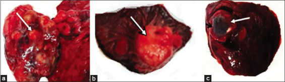
Results: Clinically, farm A and B showed the acute phase of illness and park deer showed chronic illness. Case fatality rates were 90%, 92%, and 100% in farms A, B, and C deer, respectively. Pasteurella multocida and Streptococcus pneumoniae were identified from the visceral organs of farm A deer. Farm B deer was infected with Clostridium perfringens type A. Park deer was infected with Mycobacterium tuberculosis and hydatid cyst.
Conclusion: The infectivity in farm A deer was due to stress as induced by punishing weather. The infectivity in farm B deer was due to feeding a higher volume of protein in the diet. The park C deer may optate infection from companion man and animals living around. The diseases of captive deer identified mainly were zoonotic. It needs extensive veterinary services and specialized technologies to identify these diseases, monitor the infectivity and eliminate the public health important diseases at early onset.
Keywords: deer; enterotoxaemia; mycobacterium; pasteurella; zoonosis.
Copyright: © Sultana, et al.
Figures


References
-
- Ahrar K, Imtiaz A, Iftikhar H, Nazir A. Clostridium perfringens type D enterotoximia in the Chinkara deer (Gazella bennettii) Turk. J. Vet. Anim. Sci. 2008;32(3):225–228.
-
- Audige L, Wilson P, Morris R. Disease and mortality on red deer farms in New Zealand. Vet. Rec. 2001;148(11):334–340. - PubMed
-
- Haigh J, Mackintosh C, Griffin F. Viral, parasitic and prion diseases of farmed deer and bison. Rev. Sci. Tech. 2002;21(2):219–248. - PubMed
-
- Mackintosh C, Haigh J.C, Griffin F. Bacterial diseases of farmed deer and bison. Rev. Sci. Tech. 2002;21(2):249–263. - PubMed
-
- Riaz H, Tarik M.J, Fazal M, Tanveer H, Haroon R.C, Muhammad S.A, Tasleem M.G, Aziz R. Clinico-pathologic findings of enterotoxaemia in Chinkara deer under desert conditions in Pakistan. Pak. Vet. J. 2014;34(3):400–402.
LinkOut - more resources
Full Text Sources