. 2021 Nov 18:2021:6567455.
doi: 10.1155/2021/6567455. eCollection 2021.

Effect of Magnesium on Dentinogenesis of Human Dental Pulp Cells

Affiliations

Effect of Magnesium on Dentinogenesis of Human Dental Pulp Cells

Rania M Salem et al. Int J Biomater. .

Abstract

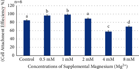
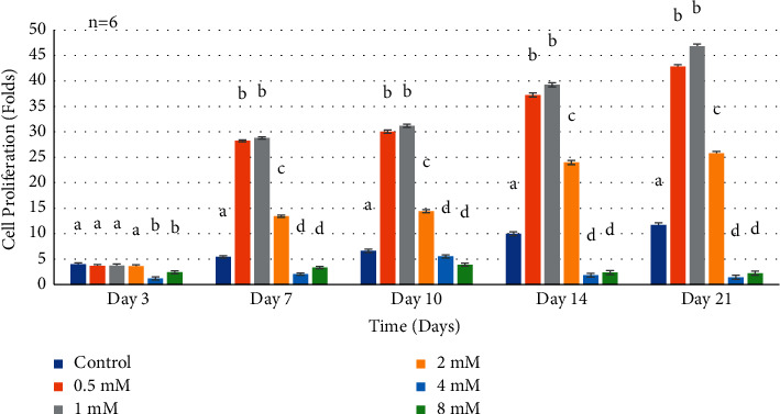
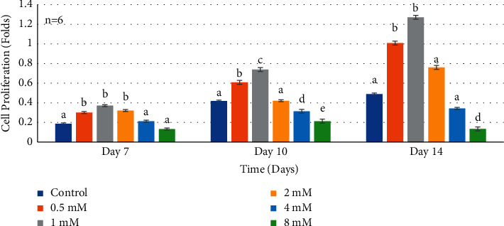
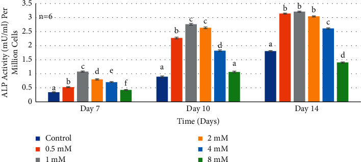
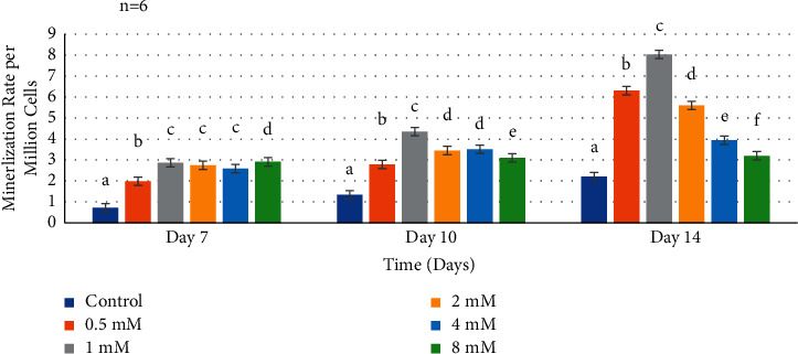
Introducing therapeutic ions into pulp capping materials has been considered a new approach for enhancing regeneration of dental tissues. However, no studies have been reported on its dentinogenic effects on human dental pulp cells (HDPCs). This study was designed to investigate the effects of magnesium (Mg2+) on cell attachment efficiency, proliferation, differentiation, and mineralization of HDPCs. HDPCs were cultured with 0.5 mM, 1 mM, 2 mM, 4 mM, and 8 mM concentrations of supplemental Mg2+ and 0 mM (control). Cell attachment was measured at 4, 8, 12, 16, and 20 hours. Cell proliferation rate was evaluated at 3, 7, 10, 14, and 21 days. Crystal violet staining was used to determine cell attachment and proliferation rate. Alkaline phosphatase (ALP) activity was assessed using the fluorometric assay at 7, 10, and 14 days. Mineralization of cultures was measured by Alizarin red staining. Statistical analysis was done using multiway analysis of variance (multiway ANOVA) with Wilks' lambda test. Higher cell attachment was shown with 0.5 mM and 1 mM at 16 hours compared to control (P < 0.0001). Cells with 0.5 mM and 1 mM supplemental Mg2+ showed significantly higher proliferation rates than control at 7, 10, 14, and 21 days (P < 0.0001). However, cell proliferation rates decreased significantly with 4 mM and 8 mM supplemental Mg2+ at 14 and 21 days (P < 0.0001). Significantly higher levels of ALP activity and mineralization were observed in 0.5 mM, 1 mM, and 2 mM supplemental Mg2+ at 10 and 14 days (P < 0.0001). However, 8 mM supplemental Mg2+ showed lower ALP activity compared to control at 14 days (P < 0.0001), while 4 mM and 8 mM supplemental Mg2+showed less mineralization compared to control (P < 0.0001). The study indicated that the optimal (0.5-2 mM) supplemental Mg2+ concentrations significantly upregulated HDPCs by enhancing cell attachment, proliferation rate, ALP activity, and mineralization. Magnesium-containing biomaterials could be considered for a future novel dental pulp-capping additive in regenerative endodontics.


Conflict of interest statement

The authors declare that they have no conflicts of interest.

Figures

Figure 1
Figure 1
Histogram showing cell attachment efficiency at 16 hours of all supplemental Mg2+ concentrations. Note: normal human dental pulp cells were cultured for 16 hours with media containing supplemental Mg2+ concentrations 0.5 mM, 1 mM, 2 mM, 4 mM, and 8 mM and 0 mM as the control group. The data are presented as means of six replicates with error bars indicating the standard deviation. Groups labeled with different letters differ statistically as compared to the control group and other study groups (P < 0.0001).
Figure 2
Figure 2
Histogram showing proliferation rates of normal human dental pulp cells in media with supplemental Mg2+ concentrations. Note: normal human dental pulp cells were cultured with media containing supplemental Mg2+concentrations 0.5 mM, 1 mM, 2 mM, 4 mM, and 8 mM and 0 mM as the control group for time periods of 3, 7, 10, 14, and 21 days. Folds were calculated by dividing the cell numbers at each time interval by the seeded cell number for each condition. Cell number was calculated by using optical density measurement results dividing the constant number (per million cells' optical density). The data are presented as means of six replicates with error bars indicating the standard deviation. Groups labeled with different letters differ statistically as compared to the control group and other study groups (P < 0.0001).
Figure 3
Figure 3
Histogram showing proliferation rates of normal human dental pulp cells in media with supplemental Mg2+ concentrations. Note: normal human dental pulp cells were cultured with media containing supplemental Mg2+concentrations 0.5 mM, 1 mM, 2 mM, 4 mM, and 8 mM and 0 mM as the control group after addition of preinductive dentinogenic media for time periods of 7, 10, and 14 days. The data are presented as means of six replicates with error bars indicating the standard deviation. Groups labeled with different letters differ statistically as compared to the control group and other study groups (P < 0.0001).
Figure 4
Figure 4
Histogram showing alkaline phosphatase activity of normal human dental pulp cells in media with supplemental Mg2+concentrations. Note: normal human dental pulp cells were cultured with media containing supplemental magnesium concentrations 0.5 mM, 1 mM, 2 mM, 4 mM, and 8 mM and 0 mM as the control group for time periods of 7, 10, and 14 days. Alkaline phosphatase activity in supernatants was normalized per million cells at each time interval. The control cells were treated with growth media without supplemental Mg2+. The data are presented as means of six replicates with error bars indicating the standard deviation. Groups labeled with different letters differ statistically as compared to the control group and other study groups (P < 0.0001).
Figure 5
Figure 5
Mineralization rate of human dental pulp cells in media with supplemental Mg2+ concentrations. Normal human dental pulp cells were cultured with media containing supplemental Mg2+concentrations 0.5 mM, 1 mM, 2 mM, 4 mM, and 8 mM and 0 mM as the control group for time periods of 7, 10, and 14 days. Mineralization of fixed cell samples was measured by spectroscopic analysis at 405 nm and normalized per million cells at each time interval. The control cells were treated with growth media without supplemental Mg2+. The data are presented as means of six replicates with error bars indicating the standard deviation. Groups labeled with different letters differ statistically as compared to the control group and other study groups (P < 0.0001).

References

    1. Flaxman D. A., Naghavi M., Lozano R., Michaud C., Ezzati M., Memish Z. A. Years lived with disability (YLDs) for 1160 sequelae of 289 diseases and injuries 1990–2010: a systematic analysis for the global burden of disease study 2010. Lancet . 2012;380(9859):2163–2196. - PMC - PubMed
    1. NICDR. Dental caries (tooth decay) in adults (age 20 to 64) 2014. http://www.nidcr.nih.gov.ezproxy.bu.edu/DataStatistics/FindDataByTopic/D... .
    1. Moussa D. G., Aparicio C. Present and future of tissue engineering scaffolds for dentin-pulp complex regeneration. Journal of Tissue Engineering and Regenerative Medicine . 2019;13(1):58–75. doi: 10.1002/term.2769. - DOI - PMC - PubMed
    1. Zhang W., Yelick P. C. Vital pulp therapy-current progress of dental pulp regeneration and revascularization. International Journal of Dentistry . 2010;2010:9. doi: 10.1155/2010/856087.856087 - DOI - PMC - PubMed
    1. Huang G. T. Pulp and dentin tissue engineering and regeneration: current progress. Regenerative Medicine . 2009;4(5):697–707. doi: 10.2217/rme.09.45. - DOI - PMC - PubMed

LinkOut - more resources