

Morphology and immunolocalization of aquaporins 1 and 9 in the agouti (Dasyprocta azarae) testis excurrent ducts
- PMID: 34840612
- PMCID: PMC8607849
- DOI: 10.1590/1984-3143-AR2021-0070
Morphology and immunolocalization of aquaporins 1 and 9 in the agouti (Dasyprocta azarae) testis excurrent ducts
Abstract
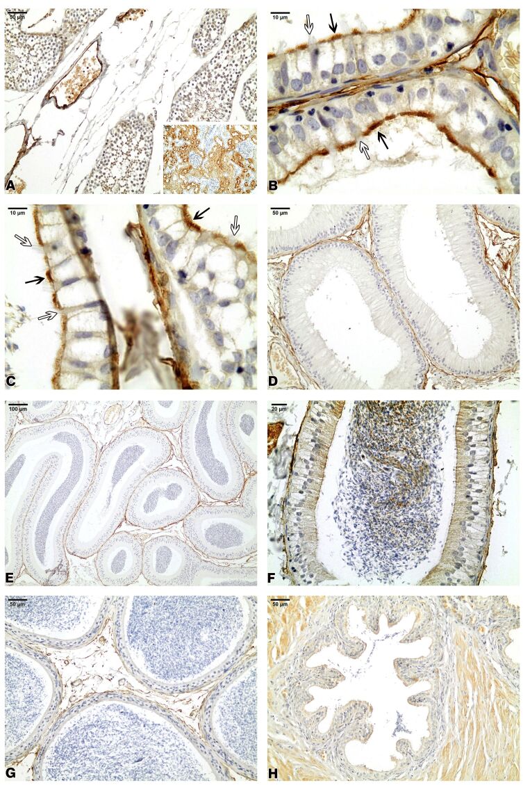
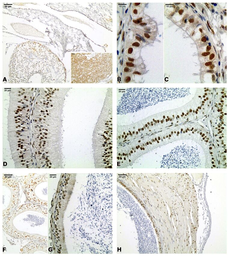
This study investigated the morphology and immunoexpression of aquaporins (AQPs) 1 and 9 in the rete testis, efferent ducts, epididymis, and vas deferens in the Azara's agouti (Dasyprocta azarae). For this purpose, ten adult sexually mature animals were used in histologic and immunohistochemical analyses. The Azara's agouti rete testis was labyrinthine and lined with simple cubic epithelium. Ciliated and non-ciliated cells were observed in the epithelium of the efferent ducts. The epididymal cellular population was composed of principal, basal, apical, clear, narrow, and halo cells. The epithelium lining of vas deferens was composed of the principal and basal cells. AQPs 1 and 9 were not expressed in the rete testis. Positive reaction to AQP1 was observed at the luminal border of non-ciliated cells of the efferent ducts, and in the peritubular stroma and blood vessels in the epididymis, and vas deferens. AQP9 was immunolocalized in the epithelial cells in the efferent ducts, epididymis and vas deferens. The morphology of Azara's agouti testis excurrent ducts is similar to that reported for other rodents such as Cuniculus paca. The immunolocalization results of the AQPs suggest that the expression of AQPs is species-specific due to differences in localization and expression when compared to studies in other mammals species. The knowledge about the expression of AQPs in Azara's agouti testis excurrent ducts is essential to support future reproductive studies on this animal, since previous studies show that AQPs may be biomarkers of male fertility and infertility.
Keywords: aquaporins; efferent ducts; epididymis; morphology; rodents.
Conflict of interest statement
Conflicts of interest: The authors have no conflict of interest to declare.
Figures


Similar articles
-
Immunolocalization of aquaporins 1, 2 and 7 in rete testis, efferent ducts, epididymis and vas deferens of adult dog.Cell Tissue Res. 2008 May;332(2):329-35. doi: 10.1007/s00441-008-0592-x. Epub 2008 Mar 14. Cell Tissue Res. 2008. PMID: 18340467
-
Aquaporin 9 (AQP9) localization in the adult dog testis excurrent ducts by immunohistochemistry.Anat Rec (Hoboken). 2007 Dec;290(12):1519-25. doi: 10.1002/ar.20611. Anat Rec (Hoboken). 2007. PMID: 17957752
-
Immunolocalization of aquaporin water channels in the domestic cat male genital tract.Reprod Domest Anim. 2014 Feb;49(1):17-26. doi: 10.1111/rda.12213. Epub 2013 Jul 5. Reprod Domest Anim. 2014. PMID: 23826797
-
Segmental and cellular expression of aquaporins in the male excurrent duct.Biochim Biophys Acta. 2006 Aug;1758(8):1025-33. doi: 10.1016/j.bbamem.2006.06.026. Epub 2006 Jul 21. Biochim Biophys Acta. 2006. PMID: 16935257 Review.
-
Thirsty business: cell, region, and membrane specificity of aquaporins in the testis, efferent ducts, and epididymis and factors regulating their expression.J Androl. 2011 Nov-Dec;32(6):565-75. doi: 10.2164/jandrol.110.012831. Epub 2011 Mar 25. J Androl. 2011. PMID: 21441426 Review.
Cited by
-
Maternal protein restriction affects the differentiation of cells in the epididymal epithelium lining of 44-day-old rats.Biol Open. 2025 Jun 15;14(6):bio060080. doi: 10.1242/bio.060080. Epub 2025 Jun 6. Biol Open. 2025. PMID: 38323644 Free PMC article.
-
Galea spixii embryos have potential to produce steroid hormones.Anim Reprod. 2023 Jan 16;19(4):e20220091. doi: 10.1590/1984-3143-AR2022-0091. eCollection 2022. Anim Reprod. 2023. PMID: 36686856 Free PMC article.
References
-
- Andonian S, Hermo L. Principal cells of the vas deferens are involved in water transport and steroid synthesis in the adult rat. J Androl. 1999;20(1):158–176. - PubMed
LinkOut - more resources
Full Text Sources