. 2021 Oct;11(5):e691-e697.
doi: 10.1212/CPJ.0000000000001084.

Multiple Spinal CSF Leaks in Spontaneous Intracranial Hypotension: Do They Exist?

Affiliations

Multiple Spinal CSF Leaks in Spontaneous Intracranial Hypotension: Do They Exist?

Wouter I Schievink et al. Neurol Clin Pract. 2021 Oct.

Abstract

Objective: To determine the frequency of multiple spinal CSF leaks in a recent group of patients with spontaneous intracranial hypotension (SIH) who were investigated with digital subtraction myelography (DSM).

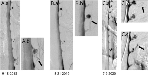
Methods: This observational study was conducted using data from a prospectively maintained data base of patients who meet the International Classification of Headache Disorders, third edition, criteria for SIH. The patient population consisted of a consecutive group of 745 patients with SIH who underwent DSM between March 2009 and February 2020. Based on the results of DSM, participants were classified according to the type and number of spinal CSF leaks.

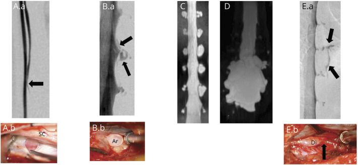
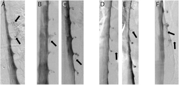
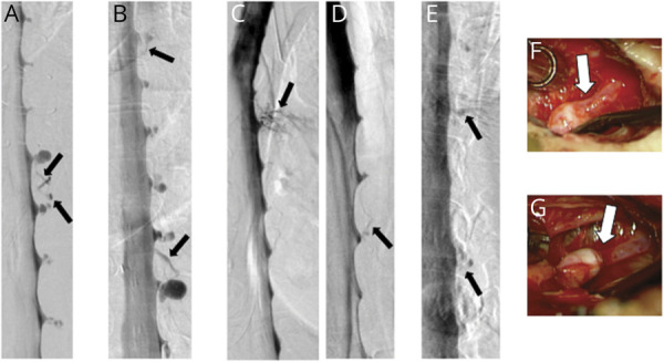
Results: Among 398 patients with SIH and extradural CSF on spinal imaging, multiplicity of CSF leaks was observed in none of 291 patients with type 1a ventral leaks and in 4 (6.2%) of 65 patients with type 1b (postero-) lateral leaks. Among 97 patients with SIH from spinal CSF-venous fistulas (type 3 leaks) who did not have extradural CSF on spinal imaging, 9 patients (9.3%) had multiple fistulas (p < 0.0001 for comparison between groups). Type 3 and type 1a or 1b CSF leaks coexisted in an additional 5 patients.

Conclusions: Among patients with SIH, multiplicity of CSF leaks was observed radiographically in none of the patients with ventral leaks, in 6% of patients with lateral leaks, and in 9% of patients with CSF-venous fistulas. These results suggest that patients with SIH can be reassured that the occurrence of multiple CSF leaks is negligible to uncommon at most, depending on the type of CSF leak.


Figures

Figure 1
Figure 1. Simplified Management Algorithm for Treatment and Imaging of Spontaneous Intracranial Hypotension (SIH)
Brain and spine imaging is used to diagnose SIH. Knowledge about the exact location of the underlying spinal CSF leak is necessary only for directed treatment with, for example, percutaneous placement of fibrin sealant, or for surgical repair, and we use digital subtraction myelography for CSF leak localization. MR = magnetic resonance.
Figure 2
Figure 2. Classification of Spontaneous Spinal CSF Leaks in Spontaneous Intracranial Hypotension
Type 1a CSF leaks are due to a ventral dural tear. Prone digital subtraction myelography (DSM) (A.a) showing location of the ventral leak (arrow) and intraoperative photograph ([A.b] posterior intradural view) showing ventral dural defect partially obscured by the spinal cord. Type 1b CSF leaks are due to a (postero)lateral dural tear. Lateral decubitus DSM (B.a) showing location of the lateral leak (arrows) and intraoperative photograph ([B.b] posterior extradural view) showing tear of the lateral thecal sac near the take-off of the nerve root sleeve with arachnoid (Ar) billowing out through the dural defect. Type 2a CSF leaks are associated with simple meningeal diverticula (C) and type 2b with dural ectasia (D). Type 3 CSF leaks are due to a CSF-venous fistula. Lateral decubitus DSM (E.a) showing location of the CSF venous fistula (arrows) and intraoperative photograph ([E.b] posterior intradural view) showing a small meningeal diverticulum (Di) transitioning into the vein (V) at the fistulous site (arrow).
Figure 3
Figure 3. Digital Subtraction Myelographies (DSMs) Demonstrating Bilateral Spontaneous Lateral (Type 1b) Dural Tears in 4 Patients
(A and B) Bilateral lateral decubitus DSMs in a patient with CSF leaks at left T10-11 and right T11-12 (arrows). (C and D) Prone DSMs in a patient with CSF leaks at left T9-10 and right T10-11 (arrows). (E and F) Bilateral lateral decubitus DSMs in a patient with bilateral CSF leaks at T10-11 (arrows). (G) Prone DSM in a patient with leaks at left T4-5 and right T3-4 (arrows).
Figure 4
Figure 4. Digital Subtraction Myelographies (DSMs) Demonstrating Multiple Spontaneous CSF-Venous Fistulas (Type 3 Leaks) in 4 Patients
(A) Lateral decubitus DSM in a patient with CSF-venous fistulas (arrows) at right T7-8 and T8-9. (B and C) Bilateral lateral decubitus DSMs in a patient with CSF-venous fistulas (arrows) at left T10-11 and right T8-9. (D and E) Lateral decubitus DSMs in a patient with CSF-venous fistulas (arrows) at left T7-8 and T9-10. (F) Lateral decubitus DSM in a patient with CSF-venous fistulas (arrows) at right T4-5 and T5-6.
Figure 5
Figure 5. Digital Subtraction Myelographies (DSMs) Demonstrating Multiple Spontaneous CSF-Venous Fistulas (Type 3 Leaks) in 4 Patients
(A) Lateral decubitus DSM in a patient with CSF-venous fistulas at left T8-9 and T9-10 (arrows). (B) Lateral decubitus DSM in a patient with CSF-venous fistulas at right T5-6 and T10-11 (arrows). (C and D) Bilateral lateral decubitus DSMs in a patient with CSF-venous fistulas at left T8-9 and right T5-6 (arrows). (E) Lateral decubitus DSM in a patient with CSF-venous fistulas at right T9-10 and T11-12 (arrows). (F and G) Corresponding intra-operative photographs confirming the presence of these 2 CSF-venous fistulas (arrows).
Figure 6
Figure 6. Digital Subtraction Myelographies (DSMs) Demonstrating 4 Spontaneous CSF-Venous Fistulas in 1 Patient, Including a De Novo Fistula
(A) Lateral decubitus DSM showing a left CSF-venous fistula at T10-11 (arrow). (B) Lateral decubitus DSM performed 8 months later now shows a left T-4-5 CSF-venous fistula (arrow) that was not present on the initial DSM despite excellent visualization of the associated meningeal diverticulum on the initial DSM. (C and D) Further lateral decubitus DSMs showing left C7-T1 (C) and T2-3 (D) CSF-venous fistulas. These levels were not as well visualized on the prior DSMs.
Figure 7
Figure 7. Coexistence of Type 1a (Ventral) or 1b (Posterolateral) CSF Leaks and CSF-Venous Fistulas (Type 3 CSF Leaks) in 5 Patients
Lateral (A) and antero-posterior (B) prone digital subtraction pyelograms (DSMs) showing a ventral CSF leak and associated CSF-venous fistula at T1-2 (arrows). C: Lateral prone DSM showing a ventral CSF leak and associated CSF-venous fistula at T11-12 (arrows). Lateral (D) and antero-posterior (E) prone DSMs showing a ventral CSF leak and associated CSF-venous fistula at T2-3 (arrows). Lateral (F) and antero-posterior (G) prone DSMs showing a ventral CSF leak and associated CSF-venous fistula at T1-2 (arrows). H: lateral decubitus DSM showing a lateral CSF leak and associated CSF-venous fistula at left T10-11 (arrows).

References

    1. Mokri B. Spontaneous cerebrospinal fluid leaks: from intracranial hypotension to cerebrospinal fluid hypovolemia—evolution of a concept. Mayo Clin Proc. 1999;74(11):1113-1123. - PubMed
    1. Schievink WI. Spontaneous spinal cerebrospinal fluid leaks and intracranial hypotension. JAMA. 2006;295(19):2286-2296. - PubMed
    1. Rahman M, Bidari SS, Quisling RG, Friedman WA. Spontaneous intracranial hypotension: dilemmas in diagnosis. Neurosurgery. 2011;69(1):4-14. - PubMed
    1. Mokri B. Spontaneous intracranial hypotension. Continuum (Minneap Minn). 2015;21(4 headache):1086-1108. - PubMed
    1. Amrhein TJ, Kranz PG. Spontaneous intracranial hypotension: imaging in diagnosis and treatment. Radiol Clin North Am. 2019;57(2):439-451. - PubMed