

Abdominal Actinomycosis Abscess Presenting as an Isolated Gastrointestinal Pseudotumor
- PMID: 34840994
- PMCID: PMC8613342
- DOI: 10.14309/crj.0000000000000672
Abdominal Actinomycosis Abscess Presenting as an Isolated Gastrointestinal Pseudotumor
Abstract
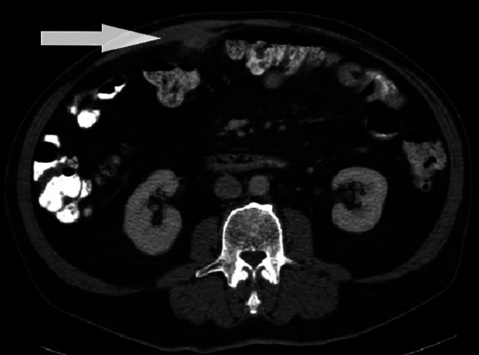
Actinomyces is a gram-positive anaerobic bacterium that is ubiquitous in nature. It typically presents as respiratory, cervicofacial, or abdominopelvic abscesses. We present a 66-year-old man with a progressive enlarging abdominal wall nodule concerning for malignancy. The patient had a negative workup, including an ultrasound-guided fine-needle aspiration and colonoscopy, with biopsy for a possible extension to the colonic wall. Diagnosis of an Actinomyces abscess was obtained through surgical resection with right hemicolectomy. He was successfully treated with a prolonged course of intravenous antibiotics. This is a rare case of an isolated abdominal wall Actinomyces abscess mimicking a gastrointestinal malignancy.
© 2021 The Author(s). Published by Wolters Kluwer Health, Inc. on behalf of The American College of Gastroenterology.
Figures


Similar articles
-
A rare finding of Actinomyces odontolyticus abdominal actinomycosis presenting as abdominal wall and pericolic pseudotumoral mass.Ann Ital Chir. 2021 Sep 27;10:S2239253X21035684. Ann Ital Chir. 2021. PMID: 34636341
-
Abdominal actinomycosis mimicking a transverse colon malignancy: a case report and review of the literature.J Med Case Rep. 2021 May 3;15(1):224. doi: 10.1186/s13256-021-02812-7. J Med Case Rep. 2021. PMID: 33934716 Free PMC article. Review.
-
[Actinomycosis Involving Chronic Pancreatitis: A Case Report with Literature Review].Korean J Gastroenterol. 2017 Mar 25;69(3):191-195. doi: 10.4166/kjg.2017.69.3.191. Korean J Gastroenterol. 2017. PMID: 28329923 Review. Korean.
-
Primary actinomycosis of anterior abdominal wall: A rare occurrence, diagnosed on fine needle aspiration cytology.Indian J Pathol Microbiol. 2019 Oct-Dec;62(4):629-630. doi: 10.4103/IJPM.IJPM_193_18. Indian J Pathol Microbiol. 2019. PMID: 31611459
-
Abdominal actinomycosis mimicking malignancy: A case report.IDCases. 2021 Aug 10;25:e01252. doi: 10.1016/j.idcr.2021.e01252. eCollection 2021. IDCases. 2021. PMID: 34430205 Free PMC article.
Cited by
-
Diffuse Large B Cell Lymphoma Raising Suspicion for an Infection: A Case Report.Cureus. 2023 Feb 7;15(2):e34750. doi: 10.7759/cureus.34750. eCollection 2023 Feb. Cureus. 2023. PMID: 36909035 Free PMC article.
-
Abdominal Actinomycosis Abscess Mimicking Malignancy: A Case Report and Review of the Literature.J Investig Med High Impact Case Rep. 2025 Jan-Dec;13:23247096251316374. doi: 10.1177/23247096251316374. J Investig Med High Impact Case Rep. 2025. PMID: 39991886 Free PMC article. Review.
-
Abdominopelvic Actinomycosis-The Diagnostic and Therapeutic Challenge of the Most Misdiagnosed Disease.Life (Basel). 2022 Mar 17;12(3):447. doi: 10.3390/life12030447. Life (Basel). 2022. PMID: 35330198 Free PMC article.
References
-
- Smego RA, Foglia G. Actinomycosis. Clin Infect Dis. 1998. 26(6):1255–3. - PubMed
-
- Piper MH, Schaberg DR, Ross JM, Shartsis JM, Orzechowski RW. Endoscopic detection and therapy of colonic actinomycosis. Am J Gastroenterol. 1992;87:1040–2. - PubMed
-
- Karaca B, Tarakci H, Tumer E, Calik S, Sen N, Sivrikoz ON. Primary abdominal wall actinomycosis. Hernia. 2015. 19, 1015–8. - PubMed
Publication types
LinkOut - more resources
Full Text Sources
