. 2021 Dec 17;24(12):103479.
doi: 10.1016/j.isci.2021.103479. Epub 2021 Nov 20.

The mRNA-LNP platform's lipid nanoparticle component used in preclinical vaccine studies is highly inflammatory

Affiliations

The mRNA-LNP platform's lipid nanoparticle component used in preclinical vaccine studies is highly inflammatory

Sonia Ndeupen et al. iScience. .

Abstract

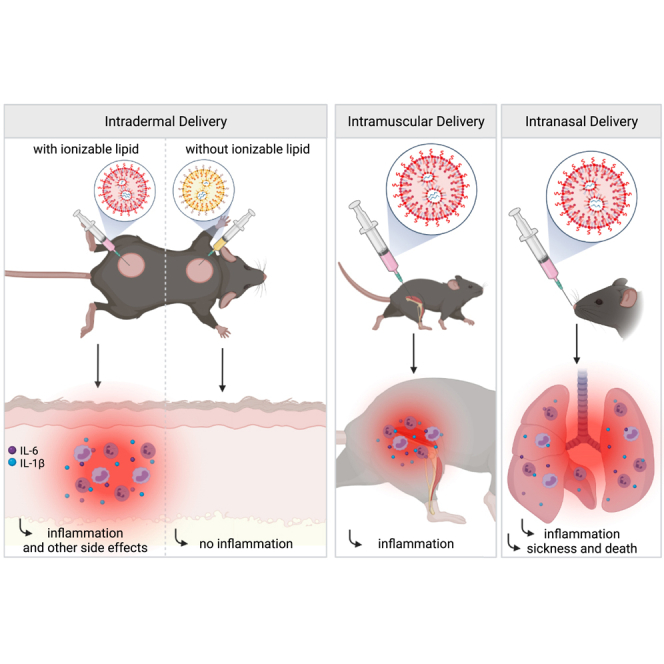
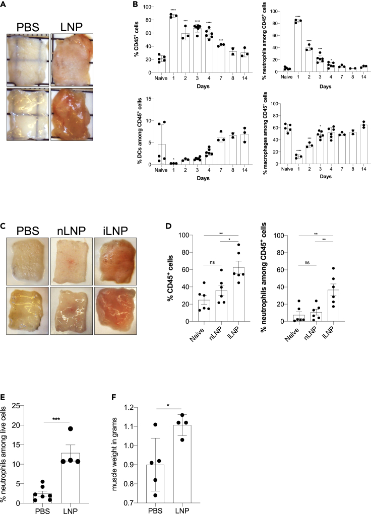
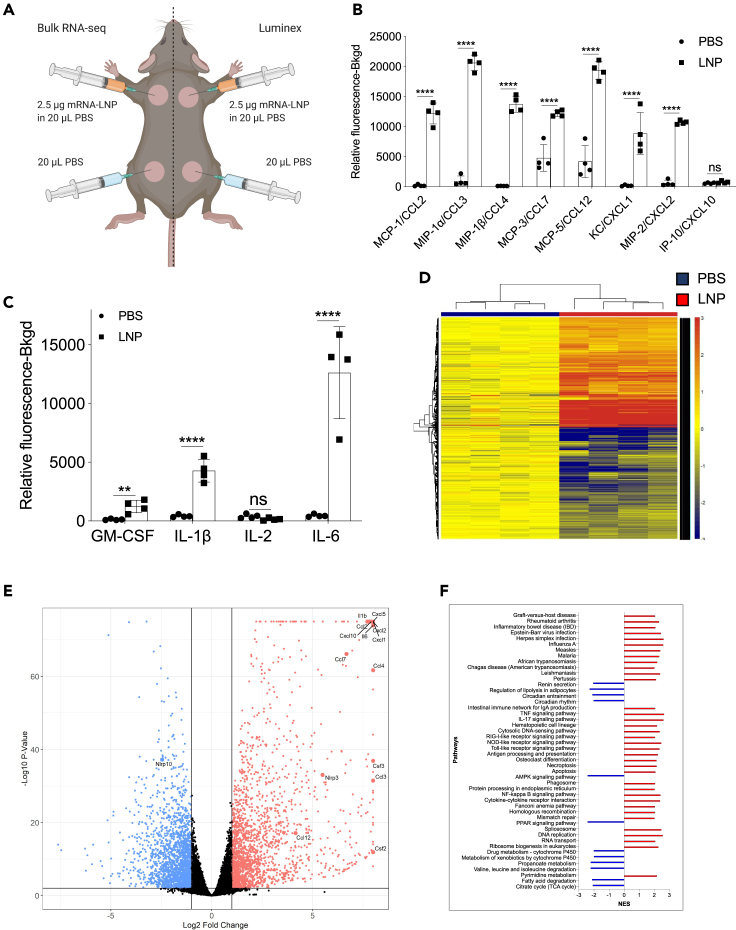
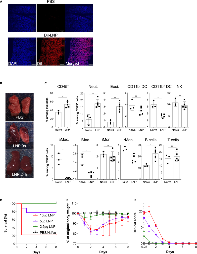
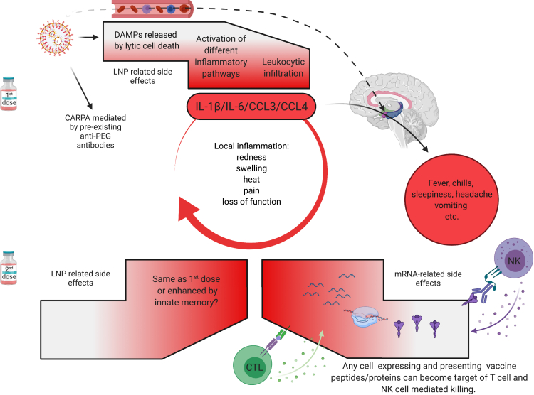
Vaccines based on mRNA-containing lipid nanoparticles (LNPs) are a promising new platform used by two leading vaccines against COVID-19. Clinical trials and ongoing vaccinations present with varying degrees of protection levels and side effects. However, the drivers of the reported side effects remain poorly defined. Here we present evidence that Acuitas' LNPs used in preclinical nucleoside-modified mRNA vaccine studies are highly inflammatory in mice. Intradermal and intramuscular injection of these LNPs led to rapid and robust inflammatory responses, characterized by massive neutrophil infiltration, activation of diverse inflammatory pathways, and production of various inflammatory cytokines and chemokines. The same dose of LNP delivered intranasally led to similar inflammatory responses in the lung and resulted in a high mortality rate, with mechanism unresolved. Thus, the mRNA-LNP platforms' potency in supporting the induction of adaptive immune responses and the observed side effects may stem from the LNPs' highly inflammatory nature.

Keywords: Biological sciences; Biotechnology; Immunology.


Conflict of interest statement

Authors declare no conflict of any sort.

Figures

None
Graphical abstract
Figure 1
Figure 1
Intradermal inoculation with LNPs induces robust inflammation (A) Intradermal inoculation with LNP induced visible levels of inflammation. Pictures were taken 24 h after PBS or LNP injection. (B) Skin samples from the mice injected with PBS or LNPs (2.5 μg/spot) were harvested at the indicated time points, analyzed by flow cytometry, and displayed as cell percentages. (C) As in A, but LNPs with (iLNP) or without (nLNP) ionizable lipids were used. Unlike iLNPs the nLNPs induced no visible signs of inflammation. (D) Skin samples from C were analyzed for leukocytic infiltration 24 h post-inoculation. (E) Muscle samples from the mice injected with PBS or 10 μg LNPs were analyzed for neutrophil infiltration 24 h post-inoculation. (F) The weight of muscle samples from E are shown. For all the charts the data were pooled from two separate experiments and displayed as percent ±SD. Each dot represents a separate animal. Student's two-tailed t test was used to determine the significance between naive and the experimental samples. ∗∗∗∗p < 0.0001, ∗∗∗p < 0.0005, ∗∗p < 0.005, ∗p < 0.05, ns = not significant. No significant differences were observed between samples harvested from naive or PBS-treated animals and are used interchangeably throughout the manuscript. See also Figure S1.
Figure 2
Figure 2
Intradermal inoculation with LNPs complexed with noncoding poly-cytosine mRNA leads to an inflammatory milieu (A) Experimental design. The mice were treated as indicated, and 24 h later, the skin samples were prepared for Luminex and bulk RNA-seq analyses. (B and C) Luminex data summarizing inflammatory chemokines and cytokines induced by the LNPs. See also Figure S2. (D) Heatmap of gene expression changes triggered by the LNPs (FDR <0.05, log2 FC > 1–4091 genes). (E) Volcano plot summarizing the up- and downregulated genes upon LNP injection. (F) GSEA analyses of the KEGG pathways and displayed as normalized enrichment score (NES). FDR<0.05. Pathways with NES less than ±2 are not displayed. N = 4.
Figure 3
Figure 3
Intranasal LNP delivery induces robust lung inflammation (A) LNP injection leads to fast and homogeneous dispersion in the lung. Animals were inoculated with PBS or 10 μg of DiI-labeled LNP. Six hours later the lungs were harvested, prepared for histology, stained with DAPI, and imaged using a confocal microscope. One representative image is shown. (B) Lungs harvested at the indicated time points from PBS, and the 10 μg LNP group were harvested and photographed. (C) Animals inoculated with 10 μg of LNP were sacrificed 9 h post inoculation and their lungs' leukocytic composition determined by flow cytometry following a published gating strategy (Yu et al., 2016). Neut. = neutrophils, Eosi. = eosinophils, DC = dendritic cells, NK = natural killer, aMac. = alveolar macrophages, iMac. = interstitial macrophages, iMon. = inflammatory monocytes, rMon. = resident monocytes. (D–F) Animals were inoculated with the indicated doses of LNP and the survival rate (D), weight (E), and clinical scores (F) recorded daily for up to 8 days. Data were pooled from two independent experiments. N = 9 for each group except PBS/naive where n = 5. For all the charts the data were pooled from at least two separate experiments and displayed as percent ±SD. Each dot represents a separate animal. Student's two-tailed t test was used to determine the significance between naive and the experimental samples. ∗∗∗p < 0.0005, ∗∗p < 0.005, ∗p < 0.05, ns = not significant. See also Figure S3.
Figure 4
Figure 4
Potential mechanism of side effects The side effects observed with the SARS-CoV-2 vaccine's first dose are likely associated with the LNPs' inflammatory properties. LNPs activate different inflammatory pathways that will lead to the production of inflammatory cytokines, such as IL-1β and IL-6, that can initiate and sustain local and systemic inflammations and side effects. The dashed line indicates the possibility that the LNPs might also diffuse from the periphery and reach any organs in the body, including CNS (hypothalamus) where they could directly induce side effects. PEG is widely used as a food and medicine additive, and many of us develop antibodies to PEG. Therefore, the LNPs' PEGylated lipids can induce CARPA in humans with preexisting PEG-specific antibodies. Humans often experience more severe side effects with the second dose. Here we posit that might be due to multiple reasons. Firstly, innate immune memory against the LNPs might form after the first vaccination and that could lead to even more robust inflammatory responses upon the second vaccination. Secondly, after the first vaccination, adaptive immune responses are formed targeting the viral protein coded by the mRNA. As such, cells (shown as red shape) expressing the viral protein-derived peptides or protein itself can become the target of CD8+ T- or NK-cell-mediated killing (ADCC), respectively. Because the LNPs could diffuse throughout the body and transfect any cell in their path with the mRNA, and the mRNA could also be further distributed through extracellular vesicles (Maugeri et al., 2019), the target population could potentially be vast and diverse.

Update of

References

    1. Abu Mouch S., Roguin A., Hellou E., Ishai A., Shoshan U., Mahamid L., Zoabi M., Aisman M., Goldschmid N., Berar Yanay N. Myocarditis following COVID-19 mRNA vaccination. Vaccine. 2021;39:3790–3793. - PMC - PubMed
    1. Achiron A., Dolev M., Menascu S., Zohar D.-N., Dreyer-Alster S., Miron S., Shirbint E., Magalashvili D., Flechter S., Givon U., et al. COVID-19 vaccination in patients with multiple sclerosis: what we have learnt by February 2021. Mult. Scler. J. 2021;27:864–870. - PMC - PubMed
    1. Alameh M.-G., Weissman D., Pardi N. Messenger RNA-based vaccines against infectious diseases. Curr. Top. Microbiol. Immunol. 2020:1–35. doi: 10.1007/82_2020_202. - DOI - PubMed
    1. Awasthi S., Hook L.M., Pardi N., Wang F., Myles A., Cancro M.P., Cohen G.H., Weissman D., Friedman H.M. Nucleoside-modified mRNA encoding HSV-2 glycoproteins C, D, and E prevents clinical and subclinical genital herpes. Sci. Immunol. 2019;4:eaaw7083. - PMC - PubMed
    1. Bernasconi V., Norling K., Gribonika I., Ong L.C., Burazerovic S., Parveen N., Schön K., Stensson A., Bally M., Larson G., et al. A vaccine combination of lipid nanoparticles and a cholera toxin adjuvant derivative greatly improves lung protection against influenza virus infection. Mucosal Immunol. 2021;14:523–536. - PubMed

LinkOut - more resources