. 2021 Jan-Dec:20:15347354211062157.
doi: 10.1177/15347354211062157.

Sporoderm-Removed Ganoderma lucidum Spore Powder May Suppress the Proliferation, Migration, and Invasion of Esophageal Squamous Cell Carcinoma Cells Through PI3K/AKT/mTOR and Erk Pathway

Affiliations

Sporoderm-Removed Ganoderma lucidum Spore Powder May Suppress the Proliferation, Migration, and Invasion of Esophageal Squamous Cell Carcinoma Cells Through PI3K/AKT/mTOR and Erk Pathway

Jingyan Liu et al. Integr Cancer Ther. 2021 Jan-Dec.

Abstract

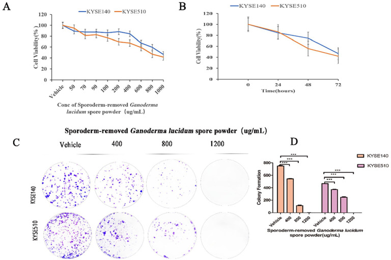
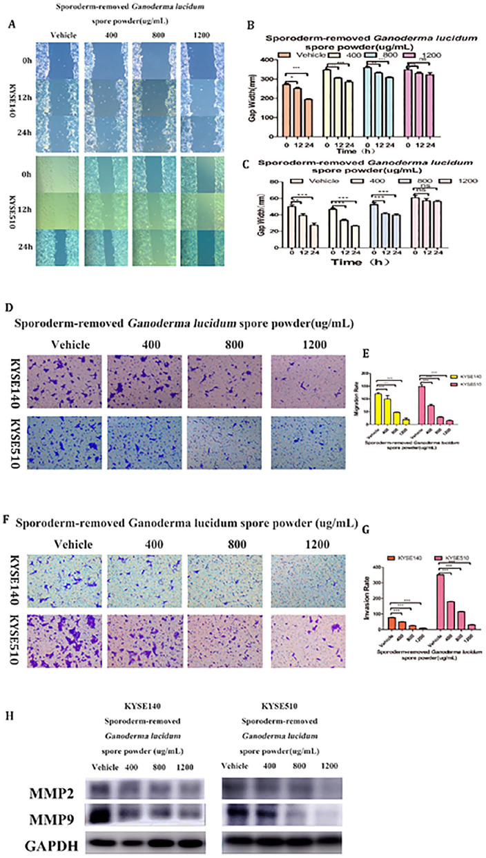
Tumor metastasis is a key factor of therapeutic failure in tumor patients, but the underlying molecular mechanism remains to be explored and novel effective curative strategies are urgently required. Emerging evidence suggests that sporoderm-removed Ganoderma lucidum spore powder can suppress tumor growth and metastasis. However, the molecular mechanisms of action remain elusive. In the present study, we investigated the effects and mechanisms of sporoderm-removed Ganoderma lucidum spore powder against esophageal squamous cell carcinomas (ESCC). The expression of MCP-1 in esophageal squamous cell carcinoma cells was detected by Western blotting. The MTS assay was used to assess the esophageal squamous cell carcinoma cells viability. The clone formation assay was used to evaluate to the proliferation ability of KYSE140 and KYSE510 cells. Apoptosis and the cell cycle were analyzed by flow cytometry. Wound healing and Transwell assays were used to analyze the migration of KYSE140 and KYSE510 cells. Invasion was also analyzed by the Transwell assay. The expressions of PI3K, AKT/p-AKT, Erk/p-Erk, JNK1, and mTOR were detected by Western blotting. We found that the MCP-1 protein was highly expressed in KYSE140 and KYSE510. In addition, sporoderm-removed Ganoderma lucidum spore powder treatment was found to inhibit esophageal squamous cell carcinoma cell proliferation, to block the cell cycle, to induce cell apoptosis and to inhibit cell migration and invasion. Finally, we found that sporoderm-removed Ganoderma lucidum spore powder decreased the expression of PI3K/AKT/mTOR and Erk signaling pathways. Taken together, these findings demonstrate that sporoderm-removed Ganoderma lucidum spore powder suppresses esophageal squamous cell carcinomas by involving MCP-1, regulated by PI3K/AKT/mTOR and Erk signal pathways.

Keywords: cell apoptosis; cell cycle; cell invasion; cell migration; cell proliferation; esophageal squamous cell carcinomas; sporoderm-removed Ganoderma lucidum spore powder.


Conflict of interest statement

Declaration of Conflicting Interests: The author(s) declared no potential conflicts of interest with respect to the research, authorship, and/or publication of this article.

Figures

Figure 1.
Figure 1.
High expression of MCP-1 in human esophageal squamous cell carcinoma cell lines.
Figure 2.
Figure 2.
Inhibition of proliferation of human esophageal squamous cell carcinoma cell lines KYSE140 and KYSE510 treated by sporoderm-removed Ganoderma lucidum spore powder. (A) KYSE140 and KYSE510 cells were treated with the indicated concentrations of sporoderm-removed Ganoderma lucidum spore powder for 72 h, cell viability was assessed using MTS assay. IC50 values were calculated using the GraphPad Prism 5.0 software. Data are presented as mean ± SD. (B) KYSE140 and KYSE510 were treated with 1000 µg/mL of sporoderm-removed Ganoderma lucidum spore powder for 24, 48, and 72 hour, respectively, cell viability was assessed using MTS assay. (C) Results of colony formation assays for KYSE140 and KYSE510 cells. ESCC cells were treated with the indicated concentrations of sporoderm-removed Ganoderma lucidum spore powder for 14 days. (D) The quantification of the cell colonies in (C) is presented as the mean percentage of viable cells (mean ± SD), averaged from 3 independent experiments, each with 3 replicates per condition.
Figure 3.
Figure 3.
MCP-1 was inhibited in KYSE140 and KYSE510 treated by sporoderm-removed Ganoderma lucidum spore powder. Western blotting analysis of MCP-1 was performed when KYSE140 and KYSE510 were treated with vehicle, 400, 800, and 1200 µg/mL of sporoderm-removed Ganoderma lucidum spore powder for 72 hours.
Figure 4.
Figure 4.
Sporoderm-removed Ganoderma lucidum spore powder arrested cell cycle in KYSE140 and KYSE510 cells. (A) KYSE140 and KYSE510 cells were treated with vehicle, 400, 800, and 1200 µg/mL of sporoderm-removed Ganoderma lucidum spore powder for 72 hours, and then stained with DAPI and subjected by FACS. (B and C) cell cycle was analyzed by Mod Fit 5.0. All data are presented as the mean ± SD. A 1-way analysis of variance, followed by a Tukey’s post-hoc test, was used to compare the different groups. *P < .05, **P < .01, ***P < .001 versus vehicle. (D) The expression of CDK1, CDK4, CDK7, p21, and p27 were examined by Western blotting. Lysates from KYSE140 and KYSE510 cell were probed with antibodies after treatment with sporoderm-removed Ganoderma lucidum spore powder for 72 hours.
Figure 5.
Figure 5.
Sporoderm-removed Ganoderma lucidum spore powder induces apoptosis in KYSE140 and KYSE510 cells. (A) KYSE140 and KYSE510 cells were treated with vehicle, 400, 800, and 1200 µg/mL of sporoderm-removed Ganoderma lucidum spore powder for 72 hours. Flow cytometry analyzed apoptotic cells stained by Annexin V and PI. (B) data are presented as the mean ± SD. A 1-way analysis of variance, followed by a Tukey’s post-hoc test, was used to compare the different groups. *P < .05, **P < .01, ***P < .001 versus vehicle. (C) Expression of the apoptosis-associated protein Fas was determined by western blotting.
Figure 6.
Figure 6.
Sporoderm-removed Ganoderma lucidum spore powder inhibited migration and invasion in KYSE140 and KYSE510 cells. (A) KYSE140 and KYSE510 cells were treated with vehicle, 400, 800, and 1200 µg/mL of sporoderm-removed Ganoderma lucidum spore powder for 72 hours. Cell migration was evaluated by the wound healing assay. Scale bar, 200 µm. (B and C) Gap Width data were presented as the mean ± SD. A 1-way analysis of variance, followed by a Tukey’s post-hoc test, was used to compare the different groups. *P < .05, **P < .01, ***P < .001 versus vehicle. (D) KYSE140 and KYSE510 cells, pretreated with vehicle, 400, 800, and 1200 µg/mL of sporoderm-removed Ganoderma lucidum spore powder for 24 hours, were plated onto the apical side of filters in serum free medium containing either vehicle or sporoderm-removed Ganoderma lucidum spore powder, medium containing 20% FBS was placed in the basolateral chamber to act as a chemoattractant for 24 hour. Cells on the bottom of the filter were stained by 0.5% crystal violet and then counted. (E) Quantification of the migrated cells in (D) was displayed on the right. The results were displayed as the mean ± SD. A 1-way analysis of variance, followed by a Tukey’s post-hoc test, was used to compare the different groups. *P < .05, **P < .01, ***P < .001 versus vehicle. (F) Before experiment, transwell chamber was covered with matrix glue. KYSE140 and KYSE510 cells, pretreated with vehicle, 400, 800, and 1200 µg/mL of sporoderm-removed Ganoderma lucidum spore powder for 24 hours, were plated onto the apical side of filters in serum free medium containing either vehicle or sporoderm-removed Ganoderma lucidum spore powder, medium containing 20% FBS was placed in the basolateral chamber to act as a chemoattractant for 24 hours. Cells on the bottom of the filter were stained by 0.5% crystal violet and then counted. (G) Quantification of the invasive cells in (F) was displayed on the right. The results were displayed as the mean ± SD. A 1-way analysis of variance, followed by a Tukey’s post-hoc test, was used to compare the different groups. *P < .05, **P < .01, ***P < .001 versus vehicle. (H) The expression of MMP2 and MMP9 that reflect cell migration and invasion was examined by Western blotting. Lysates from KYSE140 and KYSE510 cells were probed with antibodies after treatment with vehicle, 400, 800, and 1200 µg/mL of sporoderm-removed Ganoderma lucidum spore powder for 72 hour.
Figure 7.
Figure 7.
The effect of sporoderm-removed Ganoderma lucidum spore powder on PI3K/AKT/mTOR and Erk signaling pathways. The expression of PI3K, AKT, p-AKT, Erk, p-Erk, JNK1, and mTOR were assessed using the corresponding antibodies via Western blotting. The results were repeated with at least three independent experiments.

References

    1. Bray F, Ferlay J, Soerjomataram I, Siegel RL, Torre LA, Jemal A. Global cancer statistics 2018: GLOBOCAN estimates of incidence and mortality worldwide for 36 cancers in 185 countries. CA Cancer J Clin. 2018;68:394-424. doi:10.3322/caac.21492 - DOI - PubMed
    1. Chen Y, Bieerkehazhi S, Li X, Ma L, Yibulayin W, Ran J. Survivin regulates bad gene expression by binding to its promoter and modulates cell cycle and apoptosis in esophageal carcinoma cell. J Oncol. 2021;2021:1-11. doi:10.1155/2021/1384289 - DOI - PMC - PubMed
    1. Wu G, Qian Z, Guo J, et al.. Ganoderma lucidum extract induces G1 cell cycle arrest, and apoptosis in human breast cancer cells. Am J Chin Med. 2012;40:631-642. doi:10.1142/S0192415X12500474 - DOI - PubMed
    1. Li L, Guo H-J, Zhu L-Y, Zheng L, Liu X. A supercritical-CO2 extract of Ganoderma lucidum spores inhibits cholangiocarcinoma cell migration by reversing the epithelial-mesenchymal transition. Phytomedicine. 2016;23:491-497. doi:10.1016/j.phymed.2016.02.019 - DOI - PubMed
    1. Chen Y, Lv J, Li K, et al.. Sporoderm-broken spores of Ganoderma lucidum inhibit the growth of lung cancer: involvement of the Akt/mTOR signaling pathway. Nutr Cancer. 2016;68:1151-1160. doi:10.1080/01635581.2016.1208832 - DOI - PubMed

Publication types