. 2022 Dec;11(1):95-112.
doi: 10.1080/22221751.2021.2012528.

Infection and transmission of ancestral SARS-CoV-2 and its alpha variant in pregnant white-tailed deer

Affiliations

Infection and transmission of ancestral SARS-CoV-2 and its alpha variant in pregnant white-tailed deer

Konner Cool et al. Emerg Microbes Infect. 2022 Dec.

Abstract

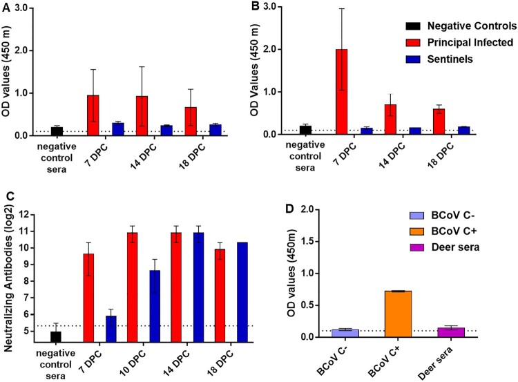
ABSTRACTSARS-CoV-2 was first reported circulating in human populations in December 2019 and has since become a global pandemic. Recent history involving SARS-like coronavirus outbreaks have demonstrated the significant role of intermediate hosts in viral maintenance and transmission. Evidence of SARS-CoV-2 natural infection and experimental infections of a wide variety of animal species has been demonstrated, and in silico and in vitro studies have indicated that deer are susceptible to SARS-CoV-2 infection. White-tailed deer (WTD) are amongst the most abundant and geographically widespread wild ruminant species in the US. Recently, WTD fawns were shown to be susceptible to SARS-CoV-2. In the present study, we investigated the susceptibility and transmission of SARS-CoV-2 in adult WTD. In addition, we examined the competition of two SARS-CoV-2 isolates, representatives of the ancestral lineage A and the alpha variant of concern (VOC) B.1.1.7 through co-infection of WTD. Next-generation sequencing was used to determine the presence and transmission of each strain in the co-infected and contact sentinel animals. Our results demonstrate that adult WTD are highly susceptible to SARS-CoV-2 infection and can transmit the virus through direct contact as well as vertically from doe to fetus. Additionally, we determined that the alpha VOC B.1.1.7 isolate of SARS-CoV-2 outcompetes the ancestral lineage A isolate in WTD, as demonstrated by the genome of the virus shed from nasal and oral cavities from principal infected and contact animals, and from the genome of virus present in tissues of principal infected deer, fetuses and contact animals.

Keywords: SARS-CoV-2; cervid; co-infection; pregnancy; susceptibility; transmission; white-tailed deer.


Conflict of interest statement

Mention of trade names or commercial products in this publication is solely for the purpose of providing specific information and does not imply recommendation or endorsement by the U.S. Department of Agriculture. USDA is an equal opportunity provider and employer. The J.A.R laboratory received support from Tonix Pharmaceuticals, Xing Technologies and Zoetis, outside of the reported work. J.A.R. is inventor on patents and patent applications on the use of antivirals and vaccines for the treatment and prevention of virus infections, owned by Kansas State University, KS, or the Icahn School of Medicine at Mount Sinai, New York. The A.G.-S. laboratory has received research support from Pfizer, Senhwa Biosciences, Kenall Manufacturing, Avimex, Johnson & Johnson, Dynavax, 7Hills Pharma, Pharmamar, ImmunityBio, Accurius, Nanocomposix, Hexamer, N-fold LLC, and Merck, outside of the reported work. A.G.-S. has consulting agreements for the following companies involving cash and/or stock: Vivaldi Biosciences, Contrafect, 7Hills Pharma, Avimex, Vaxalto, Pagoda, Accurius, Esperovax, Farmak, Applied Biological Laboratories and Pfizer, outside of the reported work. A.G.-S. is inventor on patents and patent applications on the use of antivirals and vaccines for the treatment and prevention of virus infections, owned by the Icahn School of Medicine at Mount Sinai, New York.

Figures

Figure 1.
Figure 1.
SARS-CoV-2 replication in various cervid lung cells. (A) Primary lung cells were infected with the SARS-CoV-2 USA-WA1/2020 at 0.1 MOI and cell supernatants collected at 0, 2, 4, 6 or 8 days post infection (DPI). Cell supernatants were titrated on Vero E6 cells to determine virus titres. Mean titres of at least two independent infection experiments per cell line are shown. (B) Cytopathic effect observed at 6 DPI with SARS-CoV-2 but not in mock infected white-tailed deer primary lung cells at the same time point DPI.
Figure 2.
Figure 2.
Viral shedding and viral RNA detected in tissues of SARS-CoV-2-infected white-tailed deer. RT-qPCR was performed on nasal, oropharyngeal, and rectal swabs collected from principal infected (A) and sentinel deer (B), and various tissues of deer euthanized at 4 (C) and 18 (D) days post challenge (DPC) to detect the presence of SARS-CoV-2 specific RNA. Mean (n = 2) viral RNA copy number (CN) per mL (A and B) or per mg (C and D) based on the SARS-CoV-2 nucleocapsid gene are plotted for individual animals. Asterisks (*) indicate samples with 1 out of 2 RT-qPCR reactions below the limit of detection, which is indicated by the dotted lines.
Figure 3.
Figure 3.
Histological lesions and SARS-CoV-2 antigen distribution in the upper and lower respiratory tract of principal infected white-tailed deer at 4 DPC. Rostral turbinates (A and G), olfactory neuroepithelium (B and H), trachea (C and I), bronchus (D and J) and bronchioles (E, F, K and L). At 4 DPC, in the rostral turbinates, neutrophilic rhinitis with epithelial transmigration and mixed lymphocytic and histiocytic infiltration of the subjacent lamina propria and around nasal glands were observed (A) but no viral antigen was detected (G). The olfactory neuroepithelium was histologically unremarkable (B) with no viral antigen detected (H). In the trachea, there was marked attenuation of the respiratory epithelium with loss of cilia, individual cell degeneration and necrosis, neutrophil transmigration, and accumulation of cellular debris in the lumen (C). Frequently, respiratory epithelial cells of the trachea contained intracytoplasmic viral antigen indicated by the red staining (arrows), which was also abundant in the superficial exudate (I). The bronchial mucosa was characterized by segmental attenuation of the lining respiratory epithelium with loss of cilia, degeneration/necrosis of individual epithelial cells and neutrophil and lymphocyte transmigration, and a mixed lymphocytic and histiocytic infiltrate in the edematous lamina propria (D, arrows and inset). The bronchial epithelium lining affected segments frequently contained viral antigen stained red (J, arrows). In the pulmonary parenchyma, bronchioles and blood vessels were delimited by perivascular and peribronchiolar lymphocytes, histiocytes and few neutrophils (E, arrows). Viral antigen was generally not detected (K). In bronchioles, rarely sloughed and necrotic epithelial cells and few degenerate leukocytes lodged within alveolar ducts (F, arrow) contained intracytoplasmic viral antigen indicated by the red staining (L, arrow). H&E and Fast Red, 100× total magnification.
Figure 4.
Figure 4.
Histological lesions and SARS-CoV-2 antigen distribution in the upper and lower respiratory tract of principal infected and sentinel white-tailed deer at 18 DPC. Rostral turbinates (A, F, K, P), olfactory neuroepithelium (B, G, L, Q), trachea (C, H, M, R), bronchus (D, I, N, S) and bronchioles (E, J, O, T). In principal infected deer, few lymphocytes were noted in the lamina propria of the rostral turbinates (A). The olfactory neuroepithelium was histologically unremarkable (B), and the tracheal and bronchial lamina propria were occasionally infiltrated by either dispersed or aggregates of mononuclear cells (C, D), which also encircled few bronchioles and pulmonary vessels (E). In sentinel deer, the lamina propria of rostral turbinates and sporadically subjacent to the olfactory neuroepithelium were infiltrated by mild numbers of lymphocytes and plasma cells (K, L). There was segmental erosive tracheitis with epithelial attenuation and necrosis, and intense lymphoplasmacytic and neutrophilic inflammation (M, arrows). Few mononuclear cells were noted delimiting bronchi, bronchioles and pulmonary vessels (N, O). No viral antigen was detected in respiratory tract tissues of principal infected (F–J) and sentinel (P–T) deer at 18 DPC. H&E and Fast Red, 100× total magnification.
Figure 5.
Figure 5.
Serology of SARS-CoV-2 infected deer. Detection of SARS-CoV-2 nucleocapsid protein (A), and the receptor binding domain (B) by indirect ELISA tests. The cut-off was determined by averaging the OD of negative serum + 3X the standard deviation as indicated by the dotted line. All samples with resulting OD values above this cut-off were considered positive. (C) Virus neutralizing antibodies detected in serum are shown as log2 of the reciprocal of the neutralization serum dilution. The cut-off of 1:40 is indicated by the dotted line. A–C: Negative controls were pooled deer sera (n = 4) collected from deer enrolled in a previous epizootic hemorrhagic disease virus vaccine study [23] and pre-challenge sera (-3 DPC) from the six deer enrolled in this study. (D) Sera from principal infected (n = 4) and sentinel deer (n = 2) were tested against the bovine coronavirus (BCoV) spike protein using an indirect ELISA; both, positive (C+) and negative (C−) bovine control sera were included. The cut-off was determined by averaging the OD of negative serum + 3X the standard deviation as indicated by the dotted line. A-D: Mean with standard error are shown.

Update of

References

    1. Drexler JF, Corman VM, Drosten C.. Ecology, evolution and classification of bat coronaviruses in the aftermath of SARS. Antiviral Res. 2014;101:45–56. DOI: 10.1016/j.antiviral.2013.10.013. Epub 2013/11/05. PubMed PMID: 24184128; PubMed Central PMCID: PMCPMC7113851. - DOI - PMC - PubMed
    1. de Wit E, van Doremalen N, Falzarano D, et al. SARS and MERS: recent insights into emerging coronaviruses. Nat Rev Microbiol. 2016;14(8):523–534. DOI: 10.1038/nrmicro.2016.81. Epub 2016/06/28. PubMed PMID: 27344959; PubMed Central PMCID: PMCPMC7097822. - DOI - PMC - PubMed
    1. Munnink BB, Sikkema RS, Nieuwenhuijse DF, et al. Transmission of SARS-CoV-2 on mink farms between humans and mink and back to humans. Science. 2020. DOI: 10.1126/science.abe5901. Epub 2020/11/12. PubMed PMID: 33172935 - DOI - PMC - PubMed
    1. Shi J, Wen Z, Zhong G, et al. Susceptibility of ferrets, cats, dogs, and other domesticated animals to SARS-coronavirus 2. Science. 2020;368(6494):1016–1020. DOI: 10.1126/science.abb7015. Epub 2020/04/10. PubMed PMID: 32269068; PubMed Central PMCID: PMCPMC7164390. - DOI - PMC - PubMed
    1. Halfmann PJ, Hatta M, Chiba S, et al. Transmission of SARS-CoV-2 in domestic cats. N Engl J Med. 2020;383(6):592–594. DOI: 10.1056/NEJMc2013400. Epub 2020/05/14. PubMed PMID: 32402157. - DOI - PMC - PubMed

MeSH terms

Supplementary concepts