. 2022 Jan 1;15(1):dmm049194.
doi: 10.1242/dmm.049194. Epub 2022 Jan 26.

Vangl2-environment interaction causes severe neural tube defects, without abnormal neuroepithelial convergent extension

Affiliations

Vangl2-environment interaction causes severe neural tube defects, without abnormal neuroepithelial convergent extension

Oleksandr Nychyk et al. Dis Model Mech. .

Abstract

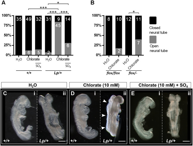
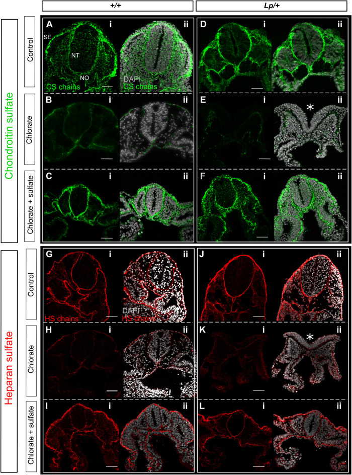
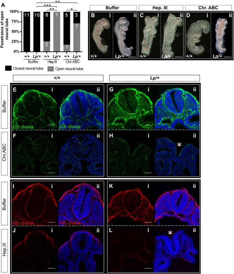
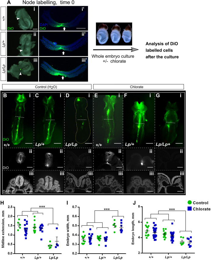
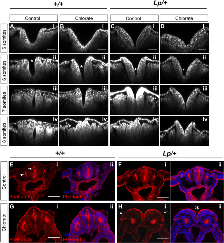
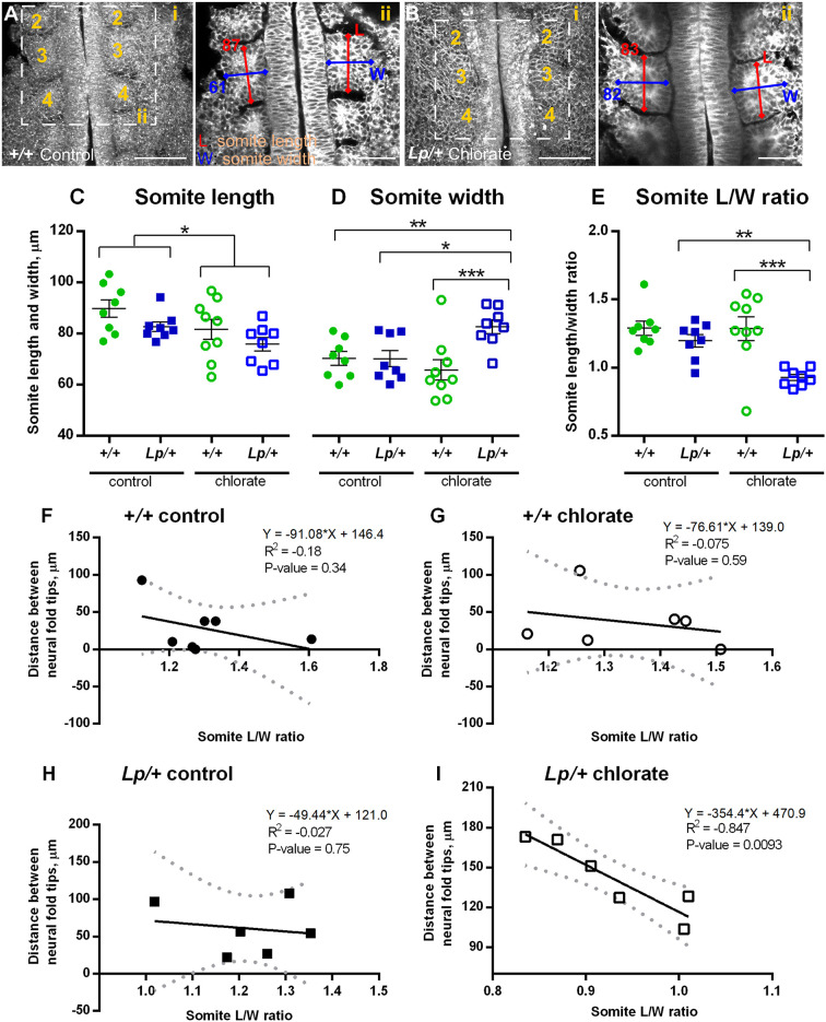
Planar cell polarity (PCP) signalling is vital for initiation of mouse neurulation, with diminished convergent extension (CE) cell movements leading to craniorachischisis, a severe neural tube defect (NTD). Some humans with NTDs also have PCP gene mutations but these are heterozygous, not homozygous as in mice. Other genetic or environmental factors may interact with partial loss of PCP function in human NTDs. We found that reduced sulfation of glycosaminoglycans interacts with heterozygosity for the Lp allele of Vangl2 (a core PCP gene), to cause craniorachischisis in cultured mouse embryos, with rescue by exogenous sulphate. We hypothesized that this glycosaminoglycan-PCP interaction may regulate CE, but, surprisingly, DiO labelling of the embryonic node demonstrates no abnormality of midline axial extension in sulfation-depleted Lp/+ embryos. Positive-control Lp/Lp embryos show severe CE defects. Abnormalities were detected in the size and shape of somites that flank the closing neural tube in sulfation-depleted Lp/+ embryos. We conclude that failure of closure initiation can arise by a mechanism other than faulty neuroepithelial CE, with possible involvement of matrix-mediated somite expansion, adjacent to the closing neural tube.

Keywords: Embryo culture; Glycosaminoglycans; Mouse; Neurulation; Planar cell polarity; Proteoglycans.


Conflict of interest statement

Competing interests The authors declare no competing or financial interests.

Figures

Fig. 1.
Fig. 1.
Expression of chondroitin sulfate (CS), heparan sulfate (HS) and Vangl2 at the Closure 1 site of wild-type embryos. (A) Schematic representation of an E8.5 mouse embryo (left), showing transverse sections through the Closure 1 region (middle) and posterior neuropore (PNP; right). A median hinge point (MHP) is prominent in the PNP, with non-bending neural plate lateral to it, whereas this is not seen at the Closure 1 site. (B,C) Immunofluorescence staining of CS (B) and HS (C) in transverse (B-i,ii,C-i,ii) and parasagittal (B-iii,iv,C-iii,iv) sections at the Closure 1 site. Both GAG chains localize to the basement membrane of NE and SE (B-i,ii,C-i,ii) (6-somite stage). CS chains are expressed at somite borders (B-iii,iv), with strong staining of HS chains at lateral junctions of SE cells (arrow in C-iii) and weak staining in NE cell membranes (arrows in C-iv). HS chains show nuclear localization in the dorsal NE and SE at the site of fusion (arrows in C-v,vi). (D) Vangl2 mRNA expression visualized by whole-mount in situ hybridization (D-i,ii) with transverse vibratome sections (D-iii-v). Transcripts are present throughout the NE, especially at the site of Closure 1 (D-i,iii,iv), and less intensely at the level of the future posterior neuropore (D-ii,v) (6-somite stage). D-ii shows a dorsal view of an open PNP, anterior to left. Vangl2 mRNA is also detected in SE (arrows in D-iii-v) and less intensely in visceral endoderm (asterisk in D-v) and paraxial mesoderm, at all levels shown. (E) Vangl2 protein is broadly expressed in NE, SE, somitic mesoderm and endoderm (6-somite stage). (F) Dorsal view of whole-mount embryo (5-somite stage) double immunostained for Vangl2 and E-cadherin (F-i,ii,iii), confirming the presence of Vangl2 in SE (F-i′,ii′,iii′). The SE and apical surface of neural plate was ‘isolated’ virtually using in-house macros. Images acquired by laser-scanning confocal microscopy using oil immersion, deconvoluted post-acquisition, and processed by single z-plane in C-iii,vi and F-i′,ii′,iii′. NE, neuroepithelium; SE, surface ectoderm; SO, somite; VE, visceral endoderm. ‘1s’, ‘2s’ and ‘3s’ indicate first, second and third somites, respectively. Scale bars: 10 µm (C-iii,vi,E-i′,ii′); 20 µm (F-i′,ii′iii′); 50 µm (B,C-i,ii,D-iii-v,E-i,ii); 100 µm (F-i,ii); 250 µm (D-i,ii).
Fig. 2.
Fig. 2.
Chlorate induces failure of Closure 1 in Vangl2Lp/+ and Vangl2flox/− embryos. (A) Experimental litters were generated by Vangl2Lp/+×Vangl2+/+ matings within the CBA/Ca-derived genetic background. Percentage of open NT (grey bar sectors) versus closed NT (black bar sectors) at the Closure 1 site was determined in cultures treated with water (control groups), 10 mM chlorate (treatment groups) or 10 mM chlorate plus sodium sulfate (rescue groups). Embryo numbers are shown on bars. Treatment with chlorate prevents Closure 1 in a significantly greater proportion of Lp/+ than +/+ embryos (P<0.001). Closure 1 is successful in 100% of water-treated controls. Co-administration of sodium sulfate (SO4) significantly reduces the Closure 1 failure seen in Lp/+ embryos treated with 10 mM chlorate (P<0.001), but not in +/+ embryos (P>0.05). (B) Chlorate significantly inhibits Closure 1 in Vangl2flox/−embryos, compared with Vangl2flox/flox, although the penetrance of Closure 1 failure after 10 mM chlorate is significantly lower in Vangl2flox/− embryos (7/18) than in Vangl2Lp/+ embryos (A; 45/54; P<0.001). (C-E) Typical embryonic morphology after 24 h culture. Vangl2+/+ embryos show completion of Closure 1 in all treatment groups (C-i,D-i,E-i). Vangl2Lp/+ water-treated (C-ii) and chlorate+sulfate-treated embryos (E-ii) also show Closure 1, whereas a chlorate-treated Vangl2Lp/+ embryo shows entirely open neural tube (arrowheads in D-ii). *P<0.05; ***P<0.001 (Chi-square test). Scale bars: 0.5 mm.
Fig. 3.
Fig. 3.
Chlorate reduces sulfation of CS and HS chains in cultured embryos. (A-L) Immunofluorescence analysis of CS (A-F) and HS (G-L) chains in embryos cultured in the presence of water (control), 10 mM chlorate, or 10 mM chlorate plus sodium sulfate. (A,D,G,J) Sections from control +/+ and Lp/+ embryos have normal distribution of CS and HS chains, which are localized to the BM underlying SE and surrounding the NT and notochord (NO); CS staining is also detected around mesenchymal cells. (B,E,H,K) Chlorate treatment dramatically reduces the staining of CS and HS chains in both genotypes and leads to failure of Closure 1 in Lp/+ embryos (asterisks in E-ii and K-ii: open NT). (C,F,I,L) Staining of CS and HS chains in +/+ and Lp/+ embryos from the rescue group (chlorate plus sulfate) appears similar to that in the control group. Note the closed NT in Lp/+ embryos. Stages shown: 12-14 somites. Scale bars: 50 µm.
Fig. 4.
Fig. 4.
Enzymatic cleavage of CS and HS chains induces Closure 1 failure in Lp/+ embryo cultures. Heparitinase III (Hep.III; to cleave HS chains, 5 U/ml) or chondroitinase ABC (Chr.ABC; to cleave CS chains, 2 U/ml) was injected into the amniotic cavity of E8.5 embryos at 0- to 5-somite stage (prior to Closure 1) at time 0, and injection was repeated after 4 h of culture. Embryos were cultured for 24 h in total. (A) All +/+ and Lp/+ embryos from the control group (enzyme buffer only) completed Closure 1. Both Hep.III and Chr.ABC significantly inhibited Closure 1, producing open NT (grey bar sectors) in Lp/+ embryos, whereas +/+ embryos remained largely unaffected. Embryo numbers are shown on bars. (B-D) Examples of embryos after culture, showing +/+ and Lp/+ buffer-treated embryos with closed NT (B-i,ii), +/+ embryos with closed NT despite treatment with enzymes (C-i,D-i), and Lp/+ embryos with failed Closure 1 after Hep.III and Chr.ABC. (E,G,I,K) Both +/+ and Lp/+ embryos from the control group (buffer) show the expected distribution of CS/HS chains after culture (compare with Fig. 3A,D,G,J, respectively). (F,H) Chr.ABC injection dramatically reduces the staining of the CS chains in both genotypes. (J,L) +/+ and Lp/+ embryos injected with Hep.III display very weak staining of HS chains. Chr.ABC does not affect HS staining, nor does Hep.III affect CS staining (Fig. S4). Asterisks indicate failed neural tube closure in Lp/+ embryos treated with Chr.ABC (H-ii) and Hep.III (L-ii). Somite stages: B-i, 12; B-ii, 13; C-i, 14; C-ii, 15; D-i, 14; D-ii, 10; E-G, 12; H, 11; I, 13; J-L, 12. *P<0.05; **P<0.01; ***P<0.001 (Chi-square test). Scale bars: 0.5 mm (B-D); 50 µm (E-L).
Fig. 5.
Fig. 5.
Chlorate prevents Closure 1 without affecting midline extension of Lp/+ embryos. (A) The node of E8.5 embryos from Vangl2Lp/+×Vangl2Lp/+ matings (0- to 5-somite stage, blind to genotype) was labelled by microinjection of the lipophilic fluorescent dye (DiO) (A-i,ii,iii). Embryos were then randomly allocated to 10 mM chlorate or water treatment and cultured for 24 h. Transverse sections through embryos at time 0 show the node and floor plate are successfully labelled with DiO in all three genotypes (A-i′,ii′,iii′). (B-G) Ventral view (rostral to the top) and transverse sections (trunk region) of DiO-injected embryos, after 24 h culture. Control (water) and chlorate-treated +/+ and Lp/+ embryos (B,C,E,F) exhibit marked midline extension of DiO-labelled cells, as detected in both whole mount and sections. Sections reveal failed Closure 1 in chlorate-treated Lp/+ embryos (F-ii,iii). In contrast, Lp/Lp embryos from both treatment groups display very limited midline extension of DiO-labelled cells (D-i,G-i), and fail in Closure 1 (D-ii,iii,G-ii,iii). Note that DiO labelling of the cranial region is non-specific, due to release of DiO into the amniotic cavity during labelling. (H) Midline extension measurements: points represent the distance DiO-labelled cells have extended along the caudal-to-rostral axis in individual embryos, with mean±s.e.m. also shown. Lp/Lp embryos exhibit significantly less midline extension than other genotypes. Chlorate-treated Lp/+ embryos do not differ from +/+ (water or chlorate) or Lp/+ (water) groups. (I,J) Embryo width (I) and length (J) measurements reveal a significantly wider and shorter body axis in Lp/Lp embryos than in other genotypes, irrespective of water/chlorate treatment. Width and length of Lp/+ embryos do not differ significantly between water and chlorate groups, nor do these values differ from +/+ embryo measurements. Data points are individual measurements, with mean±s.e.m. values also shown. *** P<0.001 (two-way ANOVA). Scale bars: 50 µm (B-ii,iii,C-ii,iii,D-ii,iii,E-ii,iii,F-ii,iii,G-ii,iii); 100 µm (A-i′,ii′,iii′); 200 µm (A-i,ii,iii); 0.5 mm (B-i,C-i,D-i,E-i,F-i,G-i).
Fig. 6.
Fig. 6.
Chlorate alters neural plate morphology but not overall F-actin distribution in cultured embryos. (A-D) Embryos were cultured for 8 h with addition of 10 mM chlorate or water as control, fixed, stained with CellMask™ and imaged using confocal microscopy for morphological analysis. Images were re-sliced in Fiji to obtain transverse sections of the Closure 1 region, at the level of the 3rd somite. +/+ (A,B) and Lp/+ (C,D) embryos with 5, 6, 7 or 8 somites are shown (i-iv for each genotype/treatment combination). Closure 1 is normally completed from the 6-somite stage onwards. In control (water-treated) +/+ embryos, the neural plate adopts an increasingly horseshoe shape, concave inwards, with fusion evident dorsally from 7 somites (A). Chlorate delays this transition in +/+ embryos, with an initially V-shaped morphology, but closure is achieved by 8 somites, when the neural tube appears largely normal (B). Water-treated Lp/+ embryos resemble chlorate-treated +/+ embryos, and achieve closure by 8 somites (C). Chlorate-treated Lp/+ embryos exhibit a persistently V-shaped neural plate with convex curvature, in which the dorsal aspects of the neural folds fail to converge and fusion fails (D). Asterisks in A-ii, B-ii and C-ii indicate sites of initial contact between neural folds. (E-H) Phalloidin staining to detect F-actin distribution in transverse sections of the Closure 1 region of embryos cultured for 24 h. F-actin is enriched at the apical surface of NE (arrow in E-i) and apically within the epithelial somites (arrowhead in E-i). Although failure of NT closure is seen in chlorate-treated Lp/+ embryos (asterisk in H-ii), the only obvious difference from +/+ (E,G) and water-treated Lp/+ embryos (F) is an apparently reduced intensity of phalloidin staining at the lateral edges of the open neural folds (arrows in H-i) (n=4 embryos each). Scale bars: 50 µm (A-D); 100 µm (E-H).
Fig. 7.
Fig. 7.
Altered somite morphology and correlation with Closure 1 delay in Lp/+ embryos after chlorate treatment. Embryos were cultured for 8 h from 0- to 5-somite stage, with addition of 10 mM chlorate or water as control, and prepared for morphological analysis as in Fig. 6A-D. (A-i,B-i) Z-projections of water-treated +/+ embryo (A-i) and chlorate-treated Lp/+ embryo (B-i), both at the 7-somite stage. Rostral is to the top; somites numbered sequentially. Images were re-sliced in Fiji (from dorsal to ventral surface) to obtain horizontal sections through the somite row. (A-ii,B-ii) Single z-planes through the Closure 1 region of the same embryos as in A-i and B-i. Length (L; rostrocaudal dimension) and width (W; mediolateral dimension) measurements were taken half way through the somite, at the level of the 3rd and 4th somites. Two somite pairs (4 individual somites) were measured per embryo. The L and W measurements (in µm) of the 3rd somite are shown for both embryos. (C-E) Somite length, width and L/W ratio in control and chlorate-treated +/+ and Lp/+ embryos, at the 6- to 7-somite stage. Individual points on the graphs are measurements averaged over 4 somites for each embryo, with mean±s.e.m. of embryo replicates. Note that somite width is significantly increased, and L/W ratio significantly reduced, in chlorate-treated Lp/+ embryos. *P<0.05; **P<0.01; ***P<0.001 (two-way ANOVA). (F-I) Linear regression analysis of distance between neural folds (µm) at the Closure 1 site and somite L/W ratios, with 95% confidence intervals (dotted lines) and a best fit line (solid lines). No significant correlation is detected for +/+ embryos, in either control (F) or chlorate (G) groups, nor for water-treated Lp/+ embryos (H). In contrast, chlorate-treated Lp/+ embryos (I) show a strong negative correlation (P=0.0093), with the greatest closure delay in embryos with the lowest somite L/W ratios (6- to 7-somite stage; n=6 in each case).

References

    1. Au, K. S., Hebert, L., Hillman, P., Baker, C., Brown, M. R., Kim, D.-K., Soldano, K., Garrett, M., Ashley-Koch, A., Lee, S.et al. (2021). Human myelomeningocele risk and ultra-rare deleterious variants in genes associated with cilium, WNT-signaling, ECM, cytoskeleton and cell migration. Sci. Rep. 11, 3639. 10.1038/s41598-021-83058-7 - DOI - PMC - PubMed
    1. Avnur, Z. and Geiger, B. (1984). Immunocytochemical localization of native chondroitin-sulfate in tissues and cultured cells using specific monoclonal antibody. Cell 38, 811-822. 10.1016/0092-8674(84)90276-9 - DOI - PubMed
    1. Bassuk, A. G., Muthuswamy, L. B., Boland, R., Smith, T. L., Hulstrand, A. M., Northrup, H., Hakeman, M., Dierdorff, J. M., Yung, C. K., Long, A.et al. (2013). Copy number variation analysis implicates the cell polarity gene glypican 5 as a human spina bifida candidate gene. Hum. Mol. Genet. 22, 1097-1111. 10.1093/hmg/dds515 - DOI - PMC - PubMed
    1. Caddy, J., Wilanowski, T., Darido, C., Dworkin, S., Ting, S. B., Zhao, Q., Rank, G., Auden, A., Srivastava, S., Papenfuss, T. A.et al. (2010). Epidermal wound repair is regulated by the planar cell polarity signaling pathway. Dev. Cell 19, 138-147. 10.1016/j.devcel.2010.06.008 - DOI - PMC - PubMed
    1. Carroll, E. A., Gerrelli, D., Gasca, S., Berg, E., Beier, D. R., Copp, A. J. and Klingensmith, J. (2003). Cordon-bleu is a conserved gene involved in neural tube formation. Dev. Biol. 262, 16-31. 10.1016/S0012-1606(03)00323-3 - DOI - PubMed

Publication types