. 2021 Dec 9;64(23):17511-17522.
doi: 10.1021/acs.jmedchem.1c01710. Epub 2021 Nov 29.

Relaxed Substrate Requirements of Sterol 14α-Demethylase from Naegleria fowleri Are Accompanied by Resistance to Inhibition

Affiliations

Relaxed Substrate Requirements of Sterol 14α-Demethylase from Naegleria fowleri Are Accompanied by Resistance to Inhibition

Tatiana Y Hargrove et al. J Med Chem. .

Abstract

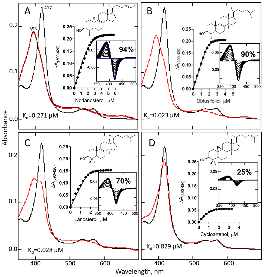
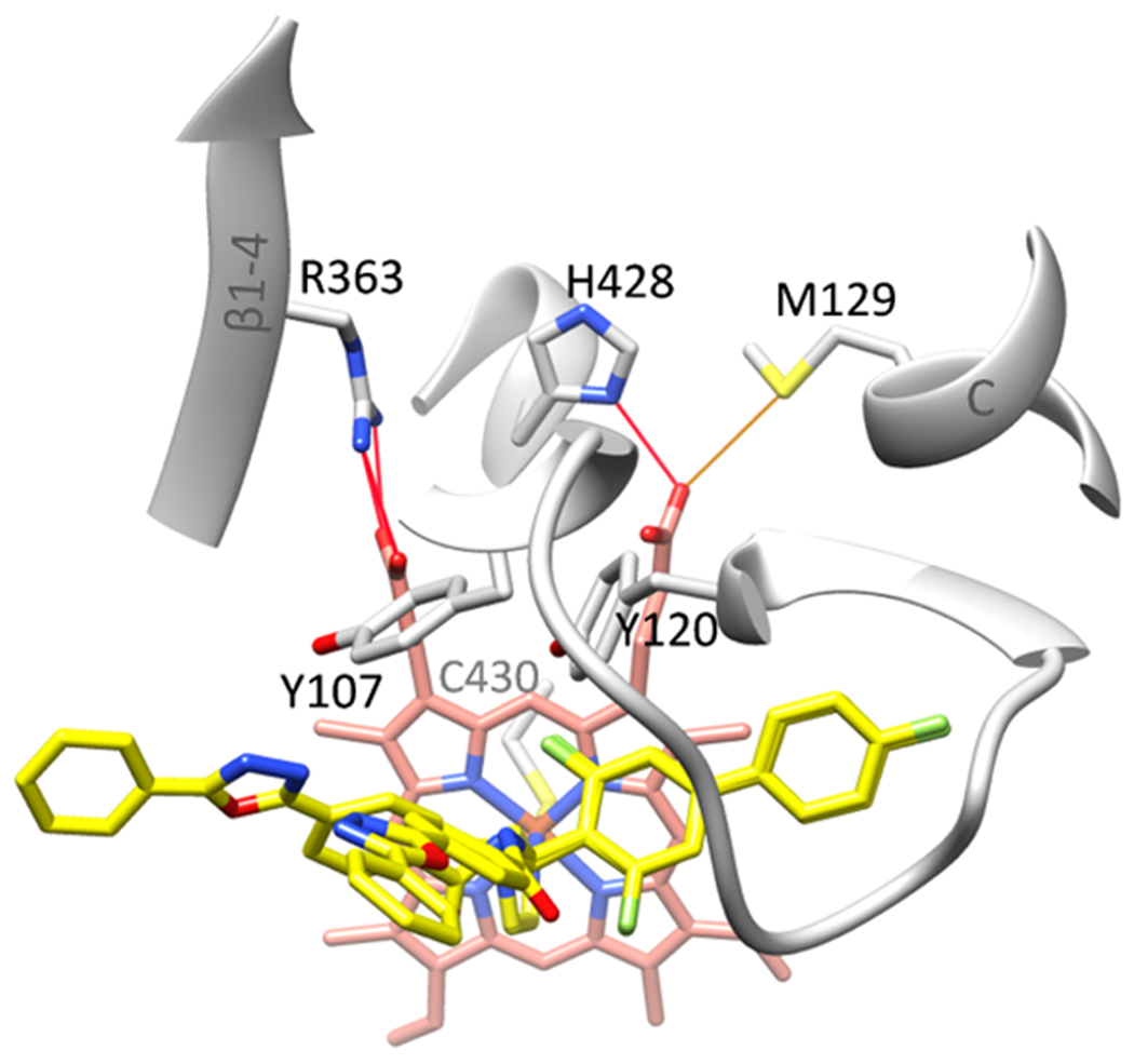
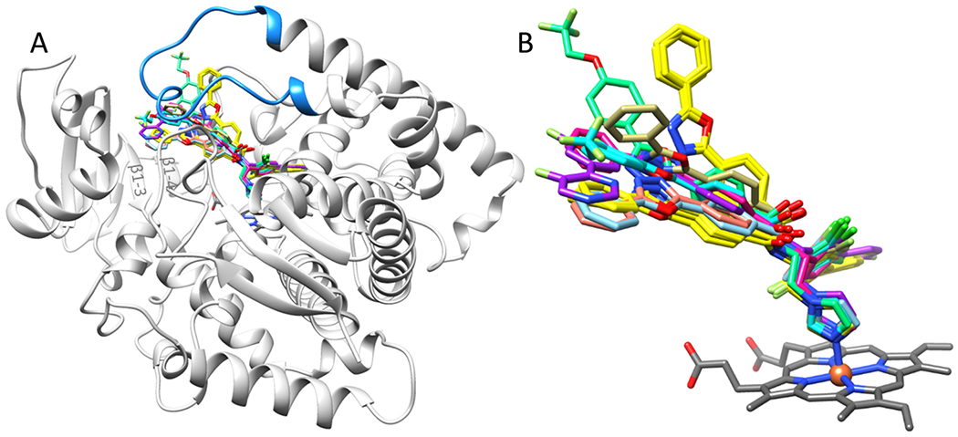
Naegleria fowleri is the protozoan pathogen that causes primary amoebic meningoencephalitis (PAM), with the death rate exceeding 97%. The amoeba makes sterols and can be targeted by sterol biosynthesis inhibitors. Here, we characterized N. fowleri sterol 14-demethylase, including catalytic properties and inhibition by clinical antifungal drugs and experimental substituted azoles with favorable pharmacokinetics and low toxicity. None of them inhibited the enzyme stoichiometrically. The highest potencies were displayed by posaconazole (IC50 = 0.69 μM) and two of our compounds (IC50 = 1.3 and 0.35 μM). Because both these compounds penetrate the brain with concentrations reaching minimal inhibitory concentration (MIC) values in an N. fowleri cellular assay, we report them as potential drug candidates for PAM. The 2.1 Å crystal structure, in complex with the strongest inhibitor, provides an explanation connecting the enzyme weaker substrate specificity with lower sensitivity to inhibition. It also provides insight into the enzyme/ligand molecular recognition process and suggests directions for the design of more potent inhibitors.


Conflict of interest statement

The authors declare no competing financial interest.

Figures

Figure 1.
Figure 1.
Sterol 14α-demethylases from different phyla. (A) Phylogenetic tree (rendered in TreeDyn 198.3). Colored leaves (top to bottom): clade Discoba phylum Heterolobosea (red); clade Discoba phylum Euglenozoa (goldenrod); clade Amoebozoa phyla Evosea (orange) and Discosea (magenta); algae (lime); plants (green); animals (blue), fungi (plum). Branch support values are displayed in %. (B) Aligned CYP51 active site areas. N. fowleri numbering is presented on the top. Residues known to be crucial for the CYP51 function and absolutely conserved across all biological domains are in red. The N. fowleri residues with the proposed specific roles (Arg108 and Met129, described later in the discussion) are marked with blue arrows. Complete sequence alignment can be found in Figure S1.
Figure 2.
Figure 2.
Absolute absorbance spectrum of purified N. fowleri CYP51. Inset: reduced carbon monoxide difference spectrum. The P450 concentration ~7 μM.
Figure 3.
Figure 3.
Spectral responses of N. fowleri CYP51 to the titration with the sterols: norlanosterol (A), obtusifoliol (B), lanosterol (C), and cycloartenol (D). Absolute absorption spectra (black) before and (red) after the titrations. Inset: the titration curves with hyperbolic fitting to the quadratic Morrison equation (A–C) or Michaelis–Menten equation (D) and the corresponding type I difference absorption spectra. The optical path length was 1 cm, and the P450 concentration was ~2 μM. Statistical parameters can be found in Figure S2.
Figure 4.
Figure 4.
Catalytic activity of N. fowleri CYP51, Michaelis–Menten curves. The experiments were performed in triplicate; the results are presented as mean ± SD. P450 concentration is 0.5 μM.
Figure 5.
Figure 5.
Spectral responses of N. fowleri CYP51 to the titration with azoles. (A) Absolute absorption spectra before (black)and after (red) the titration with ketoconazole, used as a control. Inset: the titration curve with hyperbolic fitting to the quadratic Morrison equation and the corresponding type II different absorption spectra. Panels (B) and (C) show the titration curves and difference spectral responses to the experimental compounds, VNI, VFV, and their analogues, respectively. The titration step is 0.1 μM and the optical path length is 5 cm.
Figure 6.
Figure 6.
Inhibition of N. fowleri CYP51 catalytic activity by (A) antifungal drugs (fluconazole (FLU), voriconazole (VOR), itraconazole (ITR), ketoconazole (KET), isavuconazole (ISA), and posaconazole (POS)) and experimental inhibitors: (B) VNI and analogues and (C) VFV and analogues, 1 h reaction. The experiments were performed in triplicate; the results are presented as mean ± SD. P450 concentration is 0.5 μM. Statistics for IC50 calculations is shown in Table S2.
Figure 7.
Figure 7.
F-VFV binds in the N. fowleri CYP51 active site in two conformations. (A) View from the distal P450 surface (atoms of conformer A are presented as spheres). (B) View from the upper P450 surface (atoms of conformer B are presented as spheres). The BC loop is orange; the FG loop is blue, the A’ helix is green, and the β4 hairpin is purple. Carbone atoms of the heme and inhibitor are salmon and yellow, respectively. Here and in the other figures, oxygen is red, nitrogen is blue, sulfur is yellow, and fluorine is light green or gray. (C, D) Contacts between the protein and conformers A and B, respectively, calculated in MOE using molecule A of 7RTQ. Polar interactions are in plum; hydrophobic interactions are in green. Arene–H interaction between the inhibitor distal phenyl ring of the biphenyl arm and the methyl group of the porphyrin ring D is indicated as a green dotted line. All six conformer A-specific interactions (Phe53, Pro213, Phe217, Met360, Met362, and Val468) are hydrophobic; three of the six conformer B-specific interactions (Met110, Arg111, Val113, Ile211, Ser215, and Arg226) are polar. Calculated contacts between the protein and modeled VNI/VFV analogues are shown in Figure S4.
Figure 8.
Figure 8.
Heme support in N. fowleri CYP51. Color code is the same as in Figure 7. Instead of forming H-bonds with the heme propionates, the side-chain hydroxyl of Tyr107 approaches the N3 nitrogen (2.6 Å) of the oxadiazole ring of conformer A, and the side-chain hydroxyl of Tyr120 approaches the fluorine atom of the biphenyl ring of the inhibitor (2.7 Å).
Figure 9.
Figure 9.
Substrate-preferences defining residue (F109) flips in the F-VFV-bound N. fowleri CYP51. (A) Electron density of the protein in the region around Phe109. The 2Fo-Fc map contoured at 1.5 sigma is shown as gray mesh. The ligand omit map for F-VFV is shown in Figure S6. (B) Superimposition with VFV-bound T. brucei CYP51 (light blue, the B’ helix is semitransparent, PDB ID 4g7g) and with obtusifoliol-bound I105F mutant of T. cruzi CYP51 (light brown, the B’ helix is semitransparent, PDB ID 6FMO). Orientation is similar to (A). Phe109 in N. fowleri CYP51 in trypanosomal structures aligns with Phe105, the residue that makes CYP51 prefer C4-monomethyl sterol substrates. The CYP51 signature phenylalanine (corresponds to Phe114 in N. fowleri) is also marked. As a result of the BC loop rearrangement (the flip of Phe109), the outer rings of the three-ring arm of conformer B acquire the position between the BC and FG loops.
Figure 10.
Figure 10.
Inhibitors bound in the superimposed CYP51 structures: 7RTQ (yellow), N. fowleri; 3GW9 (light blue) and 4G7G (orange), T. brucei; 4UYL (magenta) and 6CR2 (cyan—conformer A, sea green—conformer B), A. fumigatus; and 4Q2T (purple) and 4UHI (khaki) human CYP51. (A) Overall view from the lower P450 surface. The N. fowleri apoprotein ribbon and the heme are shown as markers; the FG loop is blue; and β1 strands are marked. (B) Enlarged view of the inhibitors.
Figure 11.
Figure 11.
FF-VNI and F-VFV penetrate the blood–brain barrier. Concentrations in the blood and brain after multiple doses. The compounds were given to mice by oral gavage, 25 mg/kg each 12 h for 5 days. The samples were taken 4 h after each administration. Graphs report the mean ± SEM.

References

    1. Maciver SK; Piñero JE; Lorenzo-Morales J Is Naegleria fowleri an Emerging Parasite? Trends Parasitol. 2020, 36, 19–28. - PubMed
    1. Kemble SK; Lynfield R; DeVries AS; Drehner DM; Pomputius WF III; Beach MJ; Visvesvara GS; da Silva AJ; Hill VR; Yoder JS; Xiao L; Smith KE; Danila R Fatal Naegleria fowleri Infection Acquired in Minnesota: Possible Expanded Range of a Deadly Thermophilic Organism. Clin. Infect. Dis 2012, 54, 805–809. - PubMed
    1. Linam WM; Ahmed M; Cope JR; Chu C; Visvesvara GS; da Silva AJ; Qvarnstrom Y; Green J Successful Treatment of an Adolescent with Naegleria fowleri Primary Amebic Meningoencephalitis. Pediatrics 2015, 135, e744–e748. - PMC - PubMed
    1. Gharpure R; Gleason M; Salah Z; Blackstock AJ; Hess-Homeier D; Yoder JS; Ali IKM; Collier SA; Cope JR Geographic Range of Recreational Water-Associated Primary Amebic Meningoencephalitis, United States, 1978-2018. Emerging Infect. Dis 2021, 27, 271–274. - PMC - PubMed
    1. Gharpure R; Bliton J; Goodman A; Ali IKM; Yoder J; Cope JR Epidemiology and Clinical Characteristics of Primary Amebic Meningoencephalitis Caused by Naegleria fowleri: A Global Review. Clin. Infect. Dis 2021, 73, e19–e27. - PMC - PubMed

Publication types

MeSH terms