. 2021 Nov 17;8(4):515-524.
doi: 10.3390/dermatopathology8040054.

Primary Cutaneous Gamma-Delta T Cell Lymphomas: A Case Series and Overview of the Literature

Affiliations

Primary Cutaneous Gamma-Delta T Cell Lymphomas: A Case Series and Overview of the Literature

Silvia Alberti-Violetti et al. Dermatopathology (Basel). .

Abstract

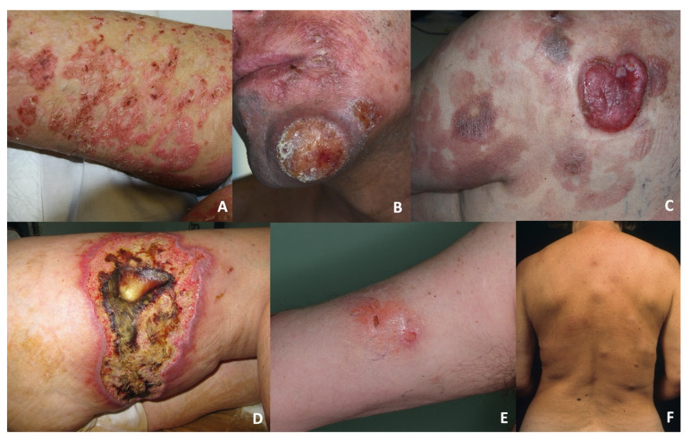
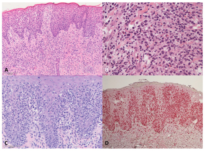
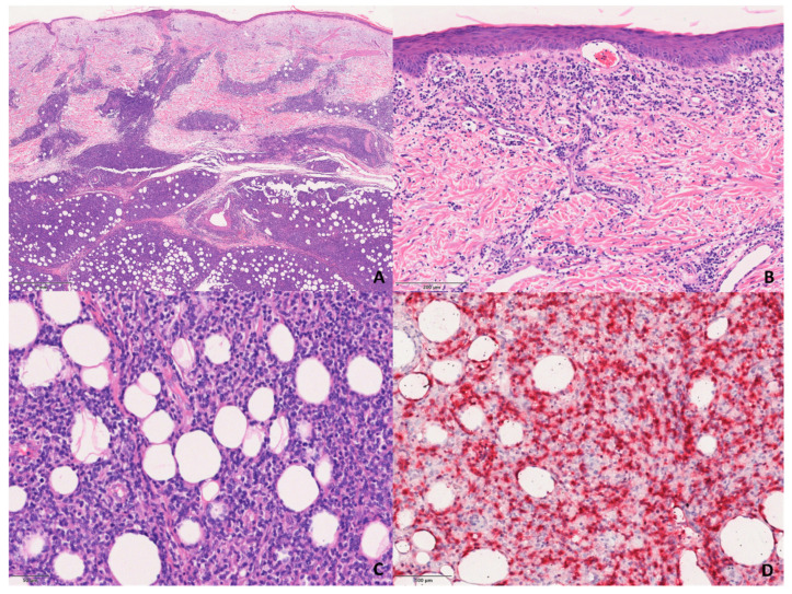
Primary cutaneous gamma-delta T cell lymphomas (PCGDTCLs) are rare and aggressive cutaneous malignancies that have been diagnostically challenging for dermopathologists and clinicians since their first published descriptions in 1991. Since then, the availability of immunostaining for T cell receptors γ and δ in formalin-fixed paraffin-embedded samples has greatly increased our knowledge of the gamma-delta phenotype by showing that it may also be present in the context of indolent entities, such as mycosis fungoides (MFs) and lymphomatoid papulosis, and this has raised questions concerning its diagnostic and prognostic implications. We here describe the histological and clinical differences between the dermo-epidermal and subcutaneous sub-groups of PCGDTCL observed in a cohort of 20 patients attending a single experienced centre, with particular focus on cases with an MF-like presentation, which are still less well defined than those of classic MF.

Keywords: cutaneous lymphoma; epidermotropic; gamma-delta; subcutaneous.


Conflict of interest statement

The authors declare no conflict of interest.

Figures

Figure 1
Figure 1
(A) A DE-GDTCL patient presenting with pagetoid reticulosis characterised by diffuse superficial patches and plaques. (B) An ulcerated tumour on the face of a DE-PCGDTCL patient. (C) A tumour associated with MF-like plaques on the back of a DE-PCGDTCL patient. (D) Pyoderma gangrenosum-like plaques on the leg of a S-PCGDTCL patient. (E) A deep erythematous plaque on the arm of an S-PCGDTCL patient. (F) Multiple deep nodules on the back of an S-PCGDTCL patient.
Figure 2
Figure 2
Histological features of the DE-GDTCLs. (A) Medium-to-large pleomorphic cells with hyper-convoluted nuclei infiltrating the epidermis and the superficial dermis (plaque). Haematoxylin and eosin (H&E), 100×. (B) Nodular infiltrate with medium-to-large pleomorphic neoplastic cells showing mitotic figures mixed with eosinophils and blasts. H&E, 400×. (C) Marked epidermotropic infiltrate of medium-sized hyperchromatic lymphocytes and some blasts. H&E, 200×. (D) TCRδ staining of the lesions shown in C. Immunophosphatase, 200×.
Figure 3
Figure 3
Histological features of the S-GDTCLs. (A) Dermal and subcutaneous involvement of the infiltrate. H&E, 200×. (B) Detail of the infiltrate in the dermis. H&E, 200×. (C) Detail of the lobular infiltrate of pleomorphic lymphocytes in subcutaneous tissue. H&E, 200×. (D) TCRδ-positive neoplastic cells in subcutaneous tissue. Immunophosphatase, 200×.

References

    1. Berti E., Willem R., Guitart J., Jaffe E.S., Kempf W., Petrella T., Robson A., Cerroni L., Torres-Cabala C.A. WHO Classification of Tumours of Haematopoietic and Lymphoid Tissues. IARC Press; Lyon, France: 2017. Primary cutaneous peripheral T-cell lymphomas, rare subtypes; pp. 397–402.
    1. Berti E., Cerri A., Cavicchini S., Delia D., Soligo D., Alessi E., Caputo R. Primary cutaneous γ/δ T-Cell Lymphoma Presenting as Disseminated Pagetoid Reticulosis. J. Investig. Dermatol. 1991;96:718–723. doi: 10.1111/1523-1747.ep12470953. - DOI - PubMed
    1. Burg G., Dummer R., Wilhelm M., Nestle F., Ott M.M., Feller A., Hefner H., Lanz U., Schwinn A., Wiede J. A Subcutaneous Delta-Positive T-Cell Lymphoma That Produces Interferon Gamma. N. Engl. J. Med. 1991;325:1078–1081. doi: 10.1056/NEJM199110103251506. - DOI - PubMed
    1. Guitart J., Martinez-Escala M.E. γδ T-cell in cutaneous and subcutaneous lymphoid infiltrates: Malignant or not? J. Cutan. Pathol. 2016;43:1242–1244. doi: 10.1111/cup.12830. - DOI - PubMed
    1. Torres-Cabala C.A., Huen A., Iyer S.P., Miranda R.N. Gamma/Delta Phenotype in Primary Cutaneous T-cell Lymphomas and Lymphoid Proliferations: Challenges for Diagnosis and Classification. Surg. Pathol. Clin. 2021;14:177–194. doi: 10.1016/j.path.2021.03.001. - DOI - PubMed