

Hair microscopy: an easy adjunct to diagnosis of systemic diseases in children
- PMID: 34843009
- PMCID: PMC8630179
- DOI: 10.1186/s42649-021-00067-6
Hair microscopy: an easy adjunct to diagnosis of systemic diseases in children
Abstract
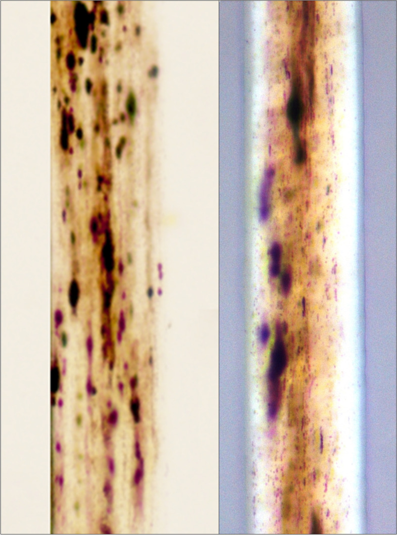
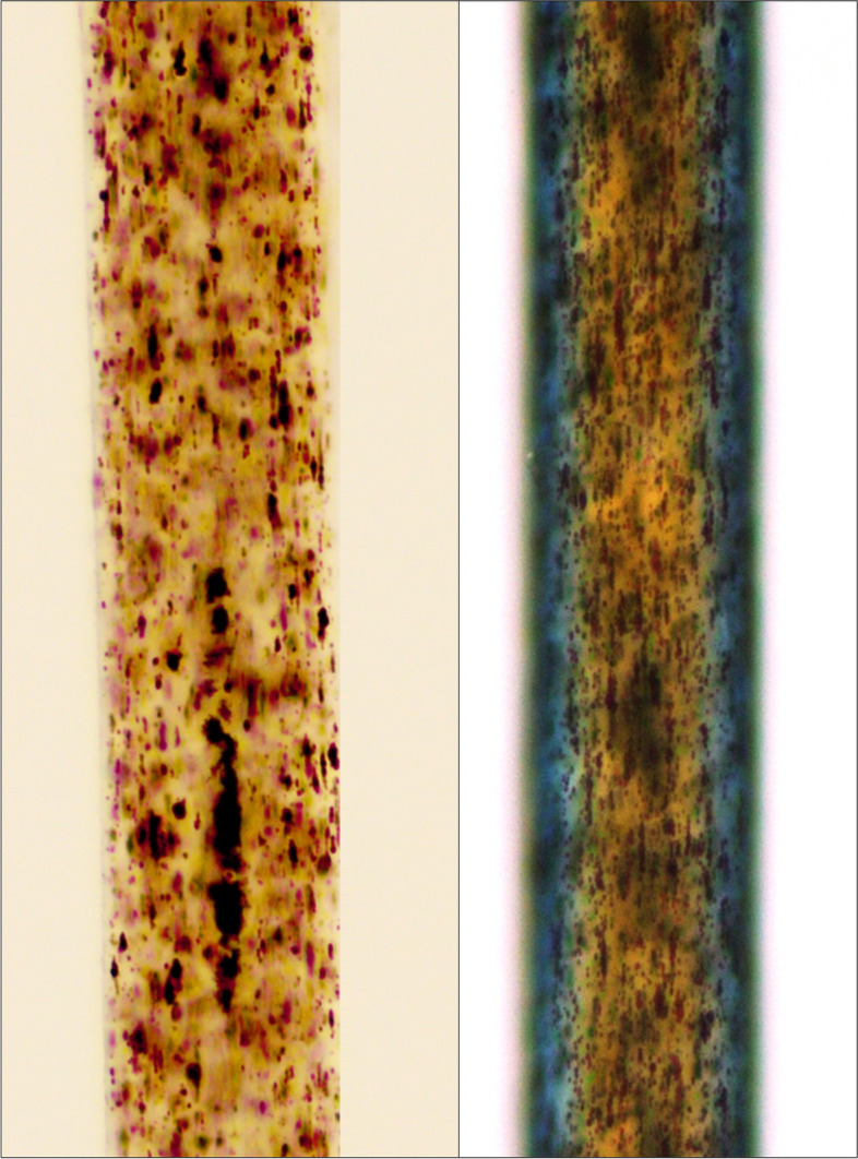
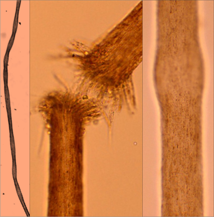
Hair, having distinct stages of growth, is a dynamic component of the integumentary system. Nonetheless, derangement in its structure and growth pattern often provides vital clues for the diagnosis of systemic diseases. Assessment of the hair structure by various microscopy techniques is, hence, a valuable tool for the diagnosis of several systemic and cutaneous disorders. Systemic illnesses like Comel-Netherton syndrome, Griscelli syndrome, Chediak Higashi syndrome, and Menkes disease display pathognomonic findings on hair microscopy which, consequently, provide crucial evidence for disease diagnosis. With minimal training, light microscopy of the hair can easily be performed even by clinicians and other health care providers which can, thus, serve as a useful tool for disease diagnosis at the patient's bedside. This is especially true for resource-constrained settings where access and availability of advanced investigations (like molecular diagnostics) is a major constraint. Despite its immense clinical utility and non-invasive nature, hair microscopy seems to be an underutilized diagnostic modality. Lack of awareness regarding the important findings on hair microscopy may be one of the crucial reasons for its underutilization. Herein, we, therefore, present a comprehensive overview of the available methods for hair microscopy and the pertinent findings that can be observed in various diseases.
Keywords: Diagnosis; Disease; Hair; Microscopy; Primary health care.
© 2021. The Author(s).
Conflict of interest statement
All authors declare to have no competing interests.
Figures
Similar articles
-
Usefulness of the skin biopsy as a tool in the diagnosis of silvery hair syndrome.Pediatr Dermatol. 2018 Nov;35(6):780-783. doi: 10.1111/pde.13624. Epub 2018 Oct 18. Pediatr Dermatol. 2018. PMID: 30338556
-
Light Microscopy and Polarized Microscopy: A Dermatological Tool to Diagnose Gray Hair Syndromes.Int J Trichology. 2017 Jan-Mar;9(1):38-41. doi: 10.4103/ijt.ijt_21_16. Int J Trichology. 2017. PMID: 28761265 Free PMC article.
-
Light microscopic examination of scalp hair samples as an aid in the diagnosis of paediatric disorders: retrospective review of more than 300 cases from a single centre.J Clin Pathol. 2005 Dec;58(12):1294-8. doi: 10.1136/jcp.2005.027581. J Clin Pathol. 2005. PMID: 16311350 Free PMC article.
-
Partial albinism with immunodeficiency: Griscelli syndrome: report of a case and review of the literature.J Am Acad Dermatol. 1998 Feb;38(2 Pt 2):295-300. doi: 10.1016/s0190-9622(98)70568-7. J Am Acad Dermatol. 1998. PMID: 9486701 Review.
-
Role of In Vivo Reflectance Confocal Microscopy in the Analysis of Melanocytic Lesions.Acta Dermatovenerol Croat. 2018 Apr;26(1):64-67. Acta Dermatovenerol Croat. 2018. PMID: 29782304 Review.
Cited by
-
Chedíak-Higashi Syndrome: Hair-to-toe spectrum.Semin Pediatr Neurol. 2024 Dec;52:101168. doi: 10.1016/j.spen.2024.101168. Epub 2024 Nov 8. Semin Pediatr Neurol. 2024. PMID: 39622608 Review.
-
Linear hair growth rates in preschool children.Pediatr Res. 2024 Jan;95(1):359-366. doi: 10.1038/s41390-023-02791-z. Epub 2023 Sep 4. Pediatr Res. 2024. PMID: 37667034 Free PMC article.
-
CBD: A Potential Lead against Hair Loss, Alopecia, and its Potential Mechanisms.Curr Drug Discov Technol. 2024;21(2):e200723218949. doi: 10.2174/1570163820666230720153607. Curr Drug Discov Technol. 2024. PMID: 37475557 Review.
-
Griscelli Syndrome: A Case Report from Pakistan, A Review of the Literature, and an Approach to Hematological Disorders Associated With Albinism.Cureus. 2025 Jun 20;17(6):e86445. doi: 10.7759/cureus.86445. eCollection 2025 Jun. Cureus. 2025. PMID: 40688996 Free PMC article.
-
A novel method for histological examination of hair follicles.Histochem Cell Biol. 2022 Jul;158(1):39-48. doi: 10.1007/s00418-022-02098-w. Epub 2022 Apr 4. Histochem Cell Biol. 2022. PMID: 35377039
References
-
- Bellon N, Hadj-Rabia S, Moulin F, Lambe C, Lezmi G, Charbit-Henrion F, Alby C, Le Saché-de Peufeilhoux L, Leclerc-Mercier S, Hadchouel A, Steffann J, Hovnanian A, Lapillonne A, Bodemer C. The challenging management of a series of 43 infants with Netherton syndrome: Unexpected complications and novel mutations. Br. J. Dermatol. 2021;184:532–537. doi: 10.1111/bjd.19265. - DOI - PubMed
Publication types
LinkOut - more resources
Full Text Sources