Review
. 2022 Feb 10:84:507-531.
doi: 10.1146/annurev-physiol-052521-121841. Epub 2021 Nov 29.

How Many Cell Types Are in the Kidney and What Do They Do?

Affiliations
Review

How Many Cell Types Are in the Kidney and What Do They Do?

Michael S Balzer et al. Annu Rev Physiol. .

Abstract

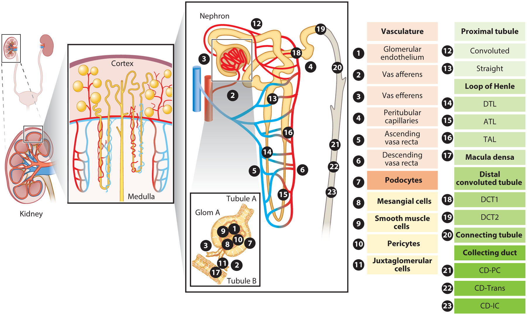
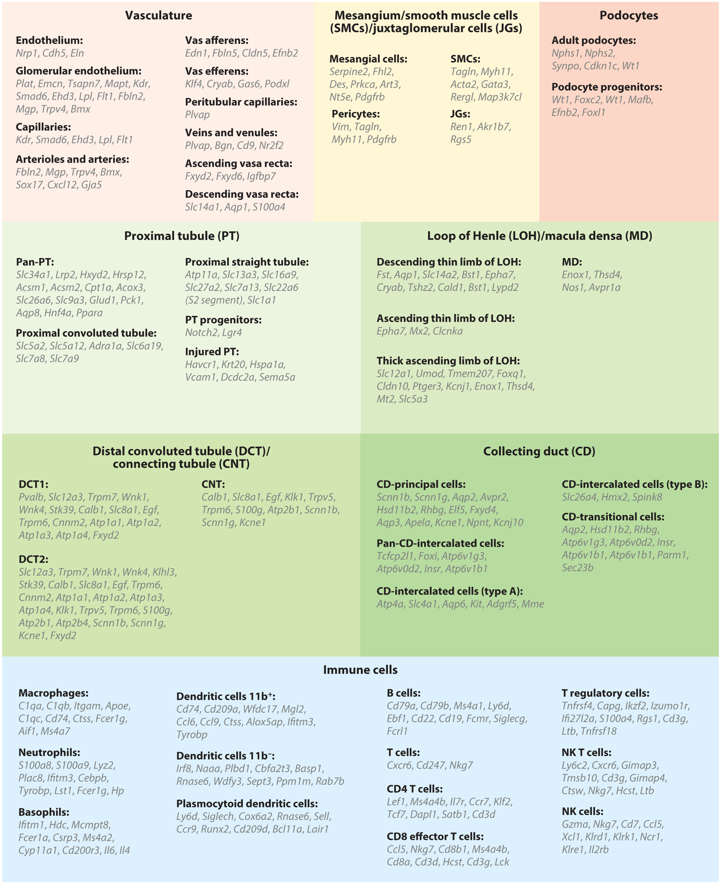
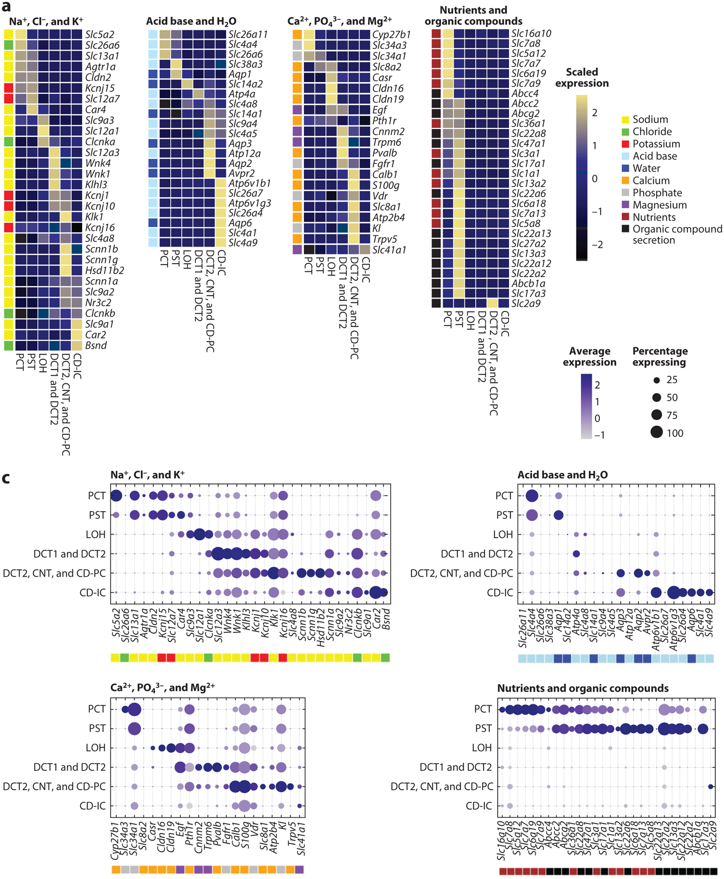
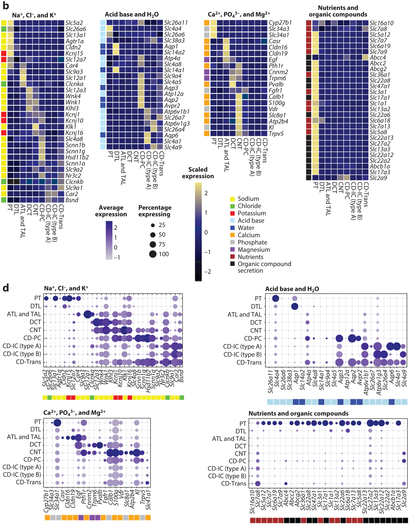
The kidney maintains electrolyte, water, and acid-base balance, eliminates foreign and waste compounds, regulates blood pressure, and secretes hormones. There are at least 16 different highly specialized epithelial cell types in the mammalian kidney. The number of specialized endothelial cells, immune cells, and interstitial cell types might even be larger. The concerted interplay between different cell types is critical for kidney function. Traditionally, cells were defined by their function or microscopical morphological appearance. With the advent of new single-cell modalities such as transcriptomics, epigenetics, metabolomics, and proteomics we are entering into a new era of cell type definition. This new technological revolution provides new opportunities to classify cells in the kidney and understand their functions.

Keywords: cell plasticity; kidney cell types; kidney function; single-cell RNA sequencing.


Figures

Figure 1
Figure 1
Overview of kidney epithelial and endothelial cell types. The various epithelial and endothelial cell types and subtypes of the kidney are numbered and annotated in the subpanel representing the functional unit of the kidney, the nephron. Kidney-resident immune cells are not included in the schematic. Abbreviations: ATL, ascending thin limb of loop of Henle; CD, collecting duct; CD-IC, collecting duct intercalated cell; CD-PC, collecting duct principal cell; CD-Trans, collecting duct transitional cell; DCT1/2, distal convoluted tubule 1 or 2; DTL, descending thin limb of loop of Henle; Glom, glomerulus; TAL, thick ascending limb of loop of Henle.
Figure 2
Figure 2
Cell type–specific marker genes of kidney epithelial, endothelial, and immune cells, as identified by single-cell RNA sequencing. Abbreviation: NK, natural killer.
Figure 3
Figure 3
Kidney-specific single-cell expression of selected genes. Selection of representative marker genes involved in metabolite handling by the different kidney tubule segments. We present two separate single-cell RNA-sequencing data sets (panels a and c and b and d, respectively) with slightly different but complementary tubular cell clustering. Subpanels are broken down by functional handling of metabolites into genes involved in the handling of sodium (Na+), chloride (Cl), and potassium (K+); acid base and water (H2O); calcium (Ca2+), phosphate (PO43−), and magnesium (Mg2+); and nutrients and organic compounds. Heatmaps (a,b) visualize row-scaled average expression (color scale), whereas dot plots (c,d) visualize the percentage of cells per cluster expressing the marker (dot size) and the average absolute gene expression (color scale). Color legends next to individual genes represent the metabolite handled by the corresponding transporter/protein. Abbreviations: ATL, ascending thin limb of LOH; CD-IC, collecting duct intercalated cell; CD-PC, collecting duct principal cell; CD-Trans, collecting duct transitional cell; CNT, connecting tubule; DCT, distal convoluted tubule; DTL, descending thin limb of LOH; LOH, loop of Henle; PCT, proximal convoluted tubule; PST, proximal straight tubule; PT, proximal tubule; TAL, thick ascending limb of LOH.
Figure 3
Figure 3
Kidney-specific single-cell expression of selected genes. Selection of representative marker genes involved in metabolite handling by the different kidney tubule segments. We present two separate single-cell RNA-sequencing data sets (panels a and c and b and d, respectively) with slightly different but complementary tubular cell clustering. Subpanels are broken down by functional handling of metabolites into genes involved in the handling of sodium (Na+), chloride (Cl), and potassium (K+); acid base and water (H2O); calcium (Ca2+), phosphate (PO43−), and magnesium (Mg2+); and nutrients and organic compounds. Heatmaps (a,b) visualize row-scaled average expression (color scale), whereas dot plots (c,d) visualize the percentage of cells per cluster expressing the marker (dot size) and the average absolute gene expression (color scale). Color legends next to individual genes represent the metabolite handled by the corresponding transporter/protein. Abbreviations: ATL, ascending thin limb of LOH; CD-IC, collecting duct intercalated cell; CD-PC, collecting duct principal cell; CD-Trans, collecting duct transitional cell; CNT, connecting tubule; DCT, distal convoluted tubule; DTL, descending thin limb of LOH; LOH, loop of Henle; PCT, proximal convoluted tubule; PST, proximal straight tubule; PT, proximal tubule; TAL, thick ascending limb of LOH.

References

    1. Bernard C 1878. Leçons sur les pheńomeǹes de la vie communs aux animaux et aux veǵet́aux. Paris: J.B. Baillier̀e
    1. Smith HW. 1953. From Fish to Philosopher. Boston: Little, Brown
    1. Smith HW. 1956. Principles of Renal Physiology. New York: Oxford Univ. Press
    1. Smith HW. 1959. The fate of sodium and water in the renal tubules. Bull. N. Y. Acad. Med 35:293–316 - PMC - PubMed
    1. Hoenig MP, Zeidel ML. 2014. Homeostasis, the milieu intérieur, and the wisdom of the nephron. Clin. J. Am. Soc. Nephrol 9:1272–81 - PMC - PubMed

Publication types

LinkOut - more resources