. 2021 Nov 29;16(11):e0260443.
doi: 10.1371/journal.pone.0260443. eCollection 2021.

Virus-infection in cochlear supporting cells induces audiosensory receptor hair cell death by TRAIL-induced necroptosis

Affiliations

Virus-infection in cochlear supporting cells induces audiosensory receptor hair cell death by TRAIL-induced necroptosis

Yushi Hayashi et al. PLoS One. .

Abstract

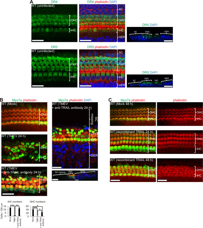
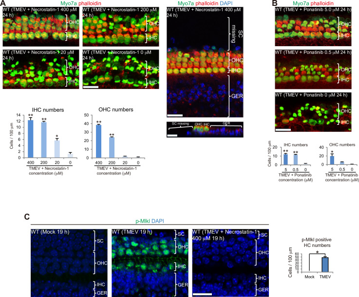
Although sensorineural hearing loss (SHL) is relatively common, its cause has not been identified in most cases. Previous studies have suggested that viral infection is a major cause of SHL, especially sudden SHL, but the system that protects against pathogens in the inner ear, which is isolated by the blood-labyrinthine barrier, remains poorly understood. We recently showed that, as audiosensory receptor cells, cochlear hair cells (HCs) are protected by surrounding accessory supporting cells (SCs) and greater epithelial ridge (GER or Kölliker's organ) cells (GERCs) against viral infections. Here, we found that virus-infected SCs and GERCs induce HC death via production of the tumour necrosis factor-related apoptosis-inducing ligand (TRAIL). Notably, the HCs expressed the TRAIL death receptors (DR) DR4 and DR5, and virus-induced HC death was suppressed by TRAIL-neutralizing antibodies. TRAIL-induced HC death was not caused by apoptosis, and was inhibited by necroptosis inhibitors. Moreover, corticosteroids, the only effective drug for SHL, inhibited the virus-induced transformation of SCs and GERCs into macrophage-like cells and HC death, while macrophage depletion also inhibited virus-induced HC death. These results reveal a novel mechanism underlying virus-induced HC death in the cochlear sensory epithelium and suggest a possible target for preventing virus-induced SHL.


Conflict of interest statement

The authors have declared that no competing interests exist.

Figures

Fig 1
Fig 1. Temporal analysis of HC death following viral infection.
(A) At 16 h after TMEV infection of the cochlear sensory epithelium, HC death was initiated in spite of the presence of very few virus-infected HCs [*P < 0.05, t-test, Mock: n = 5, TMEV: n = 4, outer HCs (OHCs), inner HCs (IHCs)]. Even at 18 h after TMEV infection when HC death was progressing, very few dsRNA-positive HCs were observed. At this time point (16 h after TMEV infection), the majority of SCs were lost by TMEV infection, which was confirmed by the computational section (see the uninfected sample). (B) Severe HC damage was observed at 24 h after TMEV infection with migration of dsRNA-positive SCs and GERCs. Most dying HCs were still negative for dsRNA. Migration of virus-infected SCs and GERCs has been reported previously [12]. (C) Ifnar1 KO and Il6 KO mice experienced SC and GERC migration to the HC layer with severe HC damage during the virus infection. (D) LPS treatment did not induce HC death with no migration of SCs and GERCs. (E) IHC and OHC numbers (counted by phalloidin staining) at 24 h of incubation with TMEV or LPS (IHC and OHC: P < 0.0001, ANOVA; *P < 0.0001, Bonferroni). Both IHCs and OHCs were reduced significantly in TMEV-infected WT cochleae (n = 3) compared with WT with mock treatment (n = 6). Even when Ifnar1 (n = 4) or Il6 (n = 3) was deficient in cochlear sensory epithelia, HC death occurred with TMEV infection similarly to that seen in WT mice, which suggested that these cytotoxic cytokines do not induce HC death. Almost all HCs survived when treated with LPS (100 ng/ml: n = 4, 1000 ng/ml: n = 4). HC death during TMEV infection was also confirmed by HC counting based on Myo7a staining (*P < 0.0001, t-test, WT mock: n = 6, WT TMEV: n = 3). These observations indicate that some kinds of secreted proteins, which were secreted when infected with TMEV, except for IFNs and IL6, but not secreted when incubated with LPS, are the cause of HC death. (F) HC stereocilia (red arrowheads) degenerated over time after activation of SCs (black arrowhead) and GERCs as macrophages (TEM; OHC1: First row of OHCs, OHC2: Second row of OHCs, OHC3: Third row of OHCs). Cytoplasmic vacuolisation has been found in many virus-infected cells [25]. (G) qRT-PCR analysis revealed that virus infection (n = 5) was more effective to upregulate Trail expression compared with LPS treatment (n = 3) or mock treatment (n = 6) at 16 h (*P < 0.05, **P < 0.01, ***P < 0.0001, t-test). Scale bars: Immunostaining, 20 μm; TEM, 10 μm. Error bars, standard errors.
Fig 2
Fig 2. TMEV infection of SCs and GERCs results in TRAIL-mediated HC death.
(A) HC expression of TRAIL receptors DR4 (green) and DR5 (green). (B) TRAIL-neutralizing antibody (5 μg/ml) attenuated HC damage (*P < 0.01, **P < 0.001, ***P < 0.0001, t-test, Mock: n = 6, TMEV: n = 4, + anti-TRAIL antibody: n = 3). While HCs were protected by anti-TRAIL antibody during TMEV infection, the death of SCs was not attenuated, indicating that the anti-TRAIL antibody has its effect directly on HCs. (C) Stereocilia of HCs was almost intact when treated with recombinant TRAIL protein (6 μg/ml) for 24 h, but long term exposure to recombinant TRAIL protein (48 h) disorganised and deformed the stereocilia. Scale bars, 20 μm. Error bars, standard errors.
Fig 3
Fig 3. Virus-infected SC- and GERC-induced HC death is not mediated by apoptosis.
(A) Cleaved caspase 3 (green) was expressed in TMEV-infected SCs and GERCs (arrowheads in sections), but not in HCs, which indicated that apoptosis did not occur in HCs. At 20 h after infection (right panel), most SCs had died by apoptosis and infected GERCs had migrated onto the HC layer, as described previously [12]. (B) Tao et al. deposited RNA sequence datasets for sorted HCs (GFP positive) and surrounding cells including SCs (GFP-negative non-HCs) from explant cultures of an Atoh1-GFP organ of Corti treated with gentamicin for 3 h in the NCBI GEO database (GSE66775) [38]. In this database, we focused on macrophage- and inflammation-correlated gene expression changes in a non-HC population including SCs to compare virus infection with aminoglycoside-related injury. Here, we extracted inflammation markers Il6 and Il1b, and macrophage markers F4/80, Mac-1, and Iba1. No significant difference was observed in the expression levels of these genes in the non-HC population, which included SCs, between the control (n = 3) and gentamicin-treated group (n = 3). Scale bars, 20 μm.
Fig 4
Fig 4. Necroptosis inhibitors supress HC damage.
(A, B) Necroptosis inhibitors necrostatin-1 (A) (IHC and OHC: P < 0.0001, ANOVA, 400 μM: n = 3, 200 μM: n = 3, 20 μM: n = 3, 0 μM: n = 4) and ponatinib (B) (IHC: P < 0.0001, OHC: P = 0.0021, ANOVA, 5 μM: n = 3, 0.5 μM: n = 4, 0 μM: n = 4) both attenuated HC death (*P < 0.001, **P < 0.0001, Bonferroni). While HCs were protected by Necrostatin-1 during TMEV infection, the death of SCs was not diminished, demonstrating that Necrostatin-1 functions directly to HCs as well as the TRAIL-neutralizing antibody. (C) p-Mlkl (green) expression in HCs induced by TMEV infection was inhibited by necrostatin-1. Thus, SCs and GERCs both induced HC necroptosis via the TRAIL-death receptor-signalling cascade. *P < 0.001, t-test, mock: n = 3, TMEV: n = 3. Scale bars, 20 μm. Error bars, standard errors.
Fig 5
Fig 5. Targeting the macrophage functions of SCs and GERCs supresses HC damage.
(A–C) Corticosteroid dexamethasone (Dex) inhibited HC damage. (A) (*P < 0.05, **P < 0.0001, t-test; TMEV + Nec (necrostatin-1): n = 3, TMEV + Dex: n = 3, TMEV: n = 4). Dex downregulated macrophage marker expression (B) (Mock: n = 3, TMEV: n = 4, TMEV + Dex: n = 5), but did not downregulate type I IFN expression at 16 h (C) (Mock: n = 5, TMEV: n = 5, TMEV + Dex: n = 5) (*P < 0.05, **P < 0.01, ***P < 0.001, ****P < 0.0001, t-test). The mechanism underlying the ability of Dex to treat SHL involves downregulation of SC and GERC macrophage functions in spite of type I IFN expression not being suppressed, which subsequently protects HCs from viral infection. (D, E) SC depletion following Clopho treatment leads to HC survival during virus infection. Clopho administration removed activated SCs as macrophages, thereby attenuating HC damage during TMEV infection (D) (*P < 0.01, **P < 0.0001, t-test, TMEV: n = 4, TMEV + Clopho: n = 3). Clopho administration to TMEV-infected explants downregulated macrophage marker expression (E) (16 h; *P < 0.05, **P < 0.01, ***P < 0.001, ****P < 0.0001, t-test, Mock: n = 3, TMEV: n = 4, TMEV + Clopho: n = 6). (F) A diagram indicating the mechanism of HC death by SCs activated as macrophage-like cells and HC protection by the glucocorticoid, macrophage-depleting agent, and necroptosis inhibitor. Scale bars = 20 μm. Error bars, standard errors.

References

    1. Davis A, McMahon CM, Pichora-Fuller KM, Russ S, Lin F, Olusanya BO, et al.. Aging and Hearing Health: The Life-course Approach. Gerontologist. 2016;56 Suppl 2:S256–67. doi: 10.1093/geront/gnw033 . - DOI - PMC - PubMed
    1. Nadol JB Jr. Hearing loss. N Engl J Med. 1993;329(15):1092–102. doi: 10.1056/NEJM199310073291507 . - DOI - PubMed
    1. Goutman JD, Elgoyhen AB, Gomez-Casati ME. Cochlear hair cells: The sound-sensing machines. FEBS Lett. 2015;589(22):3354–61. doi: 10.1016/j.febslet.2015.08.030 ; PubMed Central PMCID: PMC4641020. - DOI - PMC - PubMed
    1. Rauch SD. Clinical practice. Idiopathic sudden sensorineural hearing loss. N Engl J Med. 2008;359(8):833–40. doi: 10.1056/NEJMcp0802129 . - DOI - PubMed
    1. Schreiber BE, Agrup C, Haskard DO, Luxon LM. Sudden sensorineural hearing loss. Lancet. 2010;375(9721):1203–11. doi: 10.1016/S0140-6736(09)62071-7 . - DOI - PubMed

Publication types

MeSH terms

Substances