. 2022 Jul;142(7):1869-1881.e10.
doi: 10.1016/j.jid.2021.11.017. Epub 2021 Nov 27.

Characterization of the CpG Island Hypermethylated Phenotype Subclass in Primary Melanomas

Affiliations

Characterization of the CpG Island Hypermethylated Phenotype Subclass in Primary Melanomas

Kathleen Conway et al. J Invest Dermatol. 2022 Jul.

Abstract

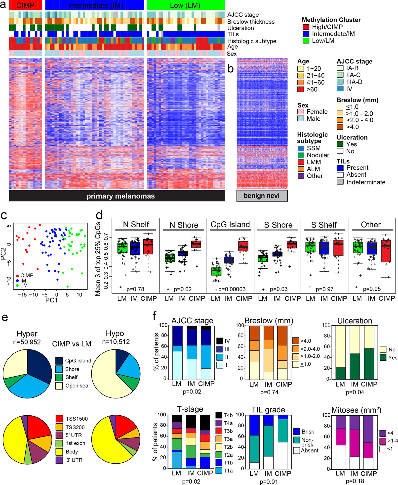
Cutaneous melanoma can be lethal even if detected at an early stage. Epigenetic profiling may facilitate the identification of aggressive primary melanomas with unfavorable outcomes. We performed clustering of whole-genome methylation data to identify subclasses that were then assessed for survival, clinical features, methylation patterns, and biological pathways. Among 89 cutaneous primary invasive melanomas, we identified three methylation subclasses exhibiting low methylation, intermediate methylation, or hypermethylation of CpG islands, known as the CpG island methylator phenotype (CIMP). CIMP melanomas occurred as early as tumor stage 1b and, compared with low-methylation melanomas, were associated with age at diagnosis ≥65 years, lentigo maligna melanoma histologic subtype, presence of ulceration, higher American Joint Committee on Cancer stage and tumor stage, and lower tumor-infiltrating lymphocyte grade (all P < 0.05). Patients with CIMP melanomas had worse melanoma-specific survival (hazard ratio = 11.84; confidence interval = 4.65‒30.20) than those with low-methylation melanomas, adjusted for age, sex, American Joint Committee on Cancer stage, and tumor-infiltrating lymphocyte grade. Genes hypermethylated in CIMP compared with those in low-methylation melanomas included PTEN, VDR, PD-L1, TET2, and gene sets related to development/differentiation, the extracellular matrix, and immunity. CIMP melanomas exhibited hypermethylation of genes important in melanoma progression and tumor immunity, and although present in some early melanomas, CIMP was associated with worse survival independent of known prognostic factors.


Conflict of interest statement

CONFLICT OF INTEREST STATEMENT

The authors state no conflict of interest.

Figures

Figure 1.
Figure 1.. DNA methylation-based primary melanoma subclasses and their characteristics.
(a) Methylation-based subclasses among 89 primary invasive melanomas denoted as low methylation (LM), intermediate methylation (IM) and high methylation or CpG island methylator phenotype (CIMP). Characteristics are indicated by bars above the heatmap. (b) Methylation profiles for 73 benign nevi. (c) Principal components analysis (PCA) illustrating three melanoma subclasses. (d) Mean β of the top 25% most variant CpGs, by subclass and genomic location. (e) Genomic locations of differentially hypermethylated or hypomethylated CpGs in the CIMP versus LM subclass; probes were restricted to those with annotation for gene, location relative to a CpG island (top) or functional region and distance from the transcription start site (TSS) (bottom). (f) Boxplots illustrating characteristics of the three methylation subclasses. Adjusted p values are from the Fisher’s exact test.
Figure 2.
Figure 2.. Survival based on methylation-based melanoma subclasses.
Kaplan-Meier plots showing melanoma-specific and all-cause survival based on the three methylation-based subclasses among (a) all melanoma patients or (b) patients without evidence of metastasis at diagnosis. Log rank p values are shown. (c) Forest plot of unadjusted hazard ratios for melanoma-specific survival, comparing the effect of methylation subclass with other prognostic and patient characteristics. The box size reflects the Cox p-value in −log10 scale. (d) Multivariable Cox proportional hazards ratios for melanoma-specific survival, adjusted for categorical variables of age (>65/≤65 yrs), sex (male/female) and AJCC stage (IA/IB, IIA/IIB/IIC, IIIA/IIIB/IIIC, IV), and TIL grade (absent/non-brisk/brisk).
Figure 3.
Figure 3.. Gene sets and pathways differentially hypomethylated in CIMP melanomas.
MethylRRA was conducted using promoter-associated CpGs/genes differentially hypomethylated (FDR p-value <0.05) between CIMP and LM subsets. (a) Bar plot showing −log10 of FDR p-values for the top enriched gene sets. Heatmap panels showing methylation in CIMP and LM tumors for selected genes involved in (b) keratinization and (c) pigmentation. The genes shown exhibited differential hypomethylation at two or more CpGs. (d) Boxplots summarizing hypomethylation of MITF and its target genes involved in pigmentation. In TCGA primary melanomas, heatmap panels show methylation and associated mRNA expression for pigmentation-related genes in CIMP vs. LM subsets identified by the 171 CpG classifier (e), and scatter plots of methylation and mRNA expression for CDK2 (f) and EDNRB (g). P-values are FDR-adjusted.
Figure 4.
Figure 4.. Selected gene sets and pathways differentially hypermethylated in CIMP melanomas.
MethylRRA analysis was conducted using promoter-associated CpGs/genes differentially hypermethylated (FDR p-value <0.05) between CIMP and LM subclasses. (a) Bar plot showing −log10 of FDR p-values for top gene sets hypermethylated in CIMP. (b) Volcano plot of CpGs hypermethylated (right) or hypomethylated (left) in promoter (red) and non-promoter (black) regions. CpGs above the broken line significant at FDR p-value <0.05. (c) Fraction of PRC2 and related gene targets defined by Ben-Porath et al (Ben-Porath et al., 2008) hypermethylated in CIMP. Heatmap panels and boxplots illustrate hypermethylation in CIMP vs. LM subclasses for VDR (d, e), PD-L1 (CD274) (g, h) and PTEN (j, k); the genes shown exhibited differential hypermethylation at two or more CpGs. Also shown are corresponding methylation and mRNA expression panels for TCGA primary melanoma subclasses defined by the 171 CpG signature, with tumors similarly ordered in both panels. Scatter plots show CpG methylation versus mRNA expression in TCGA primary melanomas for VDR (f) and PD-L1 (i), with Pearson correlations shown.
Figure 5.
Figure 5.. Hypermethylation of TET2 in the CIMP melanoma subclass.
(a) Heatmap panels showing TET2 hypermethylation in the CIMP subclass. FDR p values and CpG locations are given on the right. Methylation level is indicated by the β value, with higher methylation in red, low methylation in blue, and intermediate values shown in white. Clinical annotation is provided below the heatmap. (b) Boxplots summarizing CpG methylation differences in TET2 between melanoma subclasses. (c) Correlation of TET2 methylation of ≥0.2 β (vs. <0.2 β) with CIMP subclass status. In TCGA melanomas, (d) TET2 methylation and associated mRNA expression by subclasses defined by the 171-CpG classifier, and (e) scatter plot of TET2 methylation versus mRNA expression in primary TCGA melanomas.

Comment in

References

    1. Abe M, Ohira M, Kaneda A, Yagi Y, Yamamoto S, Kitano Y, et al. CpG island methylator phenotype is a strong determinant of poor prognosis in neuroblastomas. Cancer Res 2005;65(3):828–34. - PubMed
    1. Ableser MJ, Penuela S, Lee J, Shao Q, Laird DW. Connexin43 reduces melanoma growth within a keratinocyte microenvironment and during tumorigenesis in vivo. J Biol Chem 2014;289(3):1592–603. - PMC - PubMed
    1. AmericanCancerSociety. Cancer Facts & Figures 2020. Atlanta: American Cancer Society; 2020.
    1. Barut F, Udul P, Kokturk F, Kandemir NO, Keser SH, Ozdamar SO. Clinicopathological features and pituitary homeobox 1 gene expression in the progression and prognosis of cutaneous malignant melanoma. Kaohsiung J Med Sci 2016;32(10):494–500. - PMC - PubMed
    1. Ben-Porath I, Thomson MW, Carey VJ, Ge R, Bell GW, Regev A, et al. An embryonic stem cell-like gene expression signature in poorly differentiated aggressive human tumors. Nat Genet 2008;40(5):499–507. - PMC - PubMed

Publication types