. 2021 Nov 29;7(1):369.
doi: 10.1038/s41420-021-00761-0.

LncRNA SFTA1P mediates positive feedback regulation of the Hippo-YAP/TAZ signaling pathway in non-small cell lung cancer

Affiliations

LncRNA SFTA1P mediates positive feedback regulation of the Hippo-YAP/TAZ signaling pathway in non-small cell lung cancer

Bowen Zhu et al. Cell Death Discov. .

Abstract

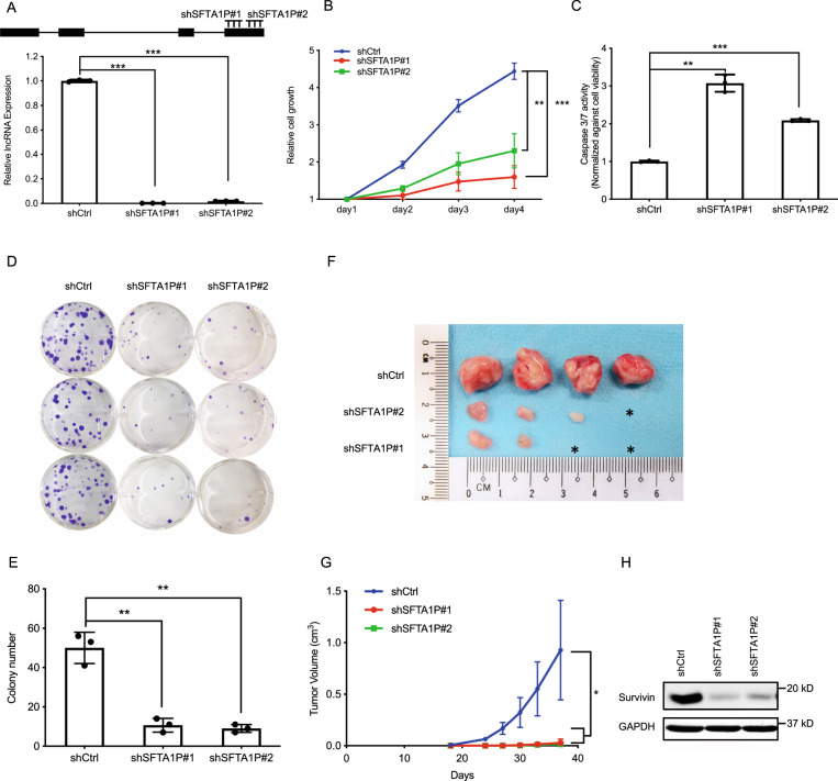
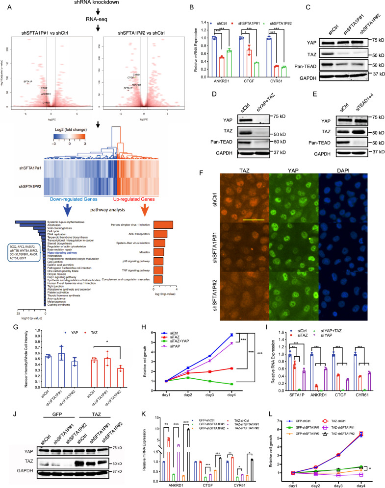
Long non-coding RNAs (lncRNAs) regulate numerous biological processes involved in both development and carcinogenesis. Hippo-YAP/TAZ signaling, a critical pathway responsible for organ size control, is often dysregulated in a variety of cancers. However, the nature and function of YAP/TAZ-regulated lncRNAs during tumorigenesis remain largely unexplored. By profiling YAP/TAZ-regulated lncRNAs, we identified SFTA1P as a novel transcriptional target and a positive feedback regulator of YAP/TAZ signaling. Using non-small cell lung cancer (NSCLC) cell lines, we show that SFTA1P is transcriptionally activated by YAP/TAZ in a TEAD-dependent manner. Functionally, knockdown of SFTA1P in NSCLC cell lines inhibited proliferation, induced programmed cell death, and compromised their tumorigenic potential. Mechanistically, SFTA1P knockdown decreased TAZ protein abundance and consequently, the expression of YAP/TAZ transcriptional targets. We provide evidence that this phenomenon could potentially be mediated via its interaction with TAZ mRNA to regulate TAZ translation. Our results reveal SFTA1P as a positive feedback regulator of Hippo-YAP/TAZ signaling, which may serve as the molecular basis for lncRNA-based therapies against YAP/TAZ-driven cancers.


Conflict of interest statement

The authors declare that they have no conflict of interest.

Figures

Fig. 1
Fig. 1. SFTA1P is transcriptionally regulated by YAP/TAZ/TEAD in NSCLC cell lines.
A A schematic illustration of the lncRNA profiling in MKN28 gastric cancer cells. SFTA1P was identified as a candidate target of YAP/TAZ. Briefly, engineered YAP/TAZ-knockout cells (YKO/TKO) were transduced with empty vector (TKO-EV or TKO) or active form of YAP (TKO-YAP or YOE)/TAZ (YKO-TAZ or TOE). RNA sequencing (n = 2 biologically independent samples) was performed to identify the differentially expressed lncRNAs with the criteria indicated. Venn diagram shows the lncRNAs that were up/downregulated upon overexpression of YAP/TAZ. Bar chart plots the relative expression of SFTA1P using Fragments Per Kilobase of transcript per Million mapped reads (FPKM). The heatmap demonstrates the relative expression of differentially expressed lncRNAs upon YAP/TAZ overexpression. The LOG2 fold-change of each lncRNA upon YAP/TAZ overexpression in two replicates were calculated and plotted. The red arrow pinpoints SFTA1P. B The expression of SFTA1P in lung cell lines was evaluated by RT-qPCR. The relative expression of SFTA1P was calculated by normalizing the SFTA1P expression in the indicated cell lines against that in the BEAS-2B cell line. Mean ± SD, n = 3 (biological replicates). C Western blot analysis of pYAP (S127), YAP, pTAZ (S89), TAZ, CYR61, and GAPDH in the indicated lung cell lines. DG Knockdown of YAP (DE), TAZ (DE), and TEADs (FG) in H1299 cells. The knockdown efficiencies were evaluated by western blot analysis in (E, G). The RNA expression of SFTA1P, ANKRD1, CTGF, and CYR61 were measured by RT-qPCR following the knockdowns of YAP and TAZ (D) or TEAD1 and TEAD4 (F), Mean ± SD, n = 4 (biological replicates). H The transcriptional activity of the regulatory sequence in the vicinity of SFTA1P TSS was assessed by dual-luciferase assay upon co-transfection of the reporter plasmids and siRNAs against YAP, TAZ, or TEADs in H1299 cells. A schematic illustration of the promoter sequence cloned into a pGL3-Basic vector was shown in the top panel: the 910 bp upstream and 2520 bp downstream sequence of the SFTA1P’s TSS (overlapping with the 1st exon, 1st intron, and the partial sequence of the 2nd exon) was examined. Mean ± SD, n = 3 (biological replicates). I Subcellular localization of SFTA1P was determined by RT-qPCR following subcellular fractionation in H1299 cells. The expression of GAPDH (cytoplasmic marker), U2 snRNA (nuclear marker), and SFTA1P were each quantified and normalized to their corresponding expression levels in the whole-cell lysate. The percentage of the expression in cytoplasmic fraction or nuclear fraction versus the summation of them were calculated and plotted. Mean ± SD, n = 3 (biological replicates). J The presence of Poly(A) tail in SFTA1P transcript was measured by RT-qPCR following reverse transcription using either random hexamer or oligo-dT. The relative expression of each gene was obtained by normalizing against the expression of beta-actin. The comparisons were calculated by normalizing the relative expression in the cDNA library synthesized using random hexamer. Mean ± SD, n = 3 (biological replicates). Statistical significance was determined using Student’s t-test, *p < 0.05, **p < 0.01, ***p < 0.001.
Fig. 2
Fig. 2. Loss of SFTA1P inhibits tumor growth in vitro and in vivo.
A Schematic annotations of SFTA1P and two shRNAs against the 4th exon of SFTA1P (top panel). The knockdown efficiencies were evaluated by RT-qPCR in H1299 cells (bottom panel). B The cell growth curve was measured by cell viability assay following shRNA-mediated silencing of SFTA1P. The relative viabilities of each treatment on day 4 were subjected to statistical analysis. Mean ± SD, n = 3 (biological replicates). C Caspase 3/7 assay measures the caspases activity upon knockdown of SFTA1P in H1299 cells. The relative caspase 3/7 activities were obtained by normalizing the raw readouts against the corresponding cell viabilities estimated by CellTiter Glo. Mean ± SD, n = 3 (biological replicates). DE Colony formation assay (D) and the corresponding quantification (E) in H1299 cell upon shRNA transduction. Mean ± SD, n = 3 (biological replicates). FG A representative image (F) and quantitative analysis (G) of the volumes of tumors formed after subcutaneously transplanting 4 × 105 H1299 cells transduced with shRNAs. Mean ± SD, n = 3 (biological replicates). H Western blot analysis of survivin expression upon the knockdown of SFTA1P. Statistical significance was determined using Student’s t-test, *p < 0.05, **p < 0.01, ***p < 0.001.
Fig. 3
Fig. 3. SFTA1P mediates a positive feedback regulation of the Hippo-YAP/TAZ signaling pathway by targeting TAZ.
A RNA-seq analysis revealed the differentially expressed genes following the knockdown of SFTA1P in H1299 cells. Volcano plots show the LOG2 fold-changes in gene expression (x-axis) and the −Log10 of the Benjamini-Hochberg adjusted P-values (y-axis) between the cells treated with shSFTA1P#1 (left panel) or shSFTA1P#2 (right panel) versus shCtrl. n = 2 (biological replicates). SFTA1P and transcriptional targets of YAP/TAZ were highlighted in black and green, respectively. Heatmap shows deferentially expressed genes following the shRNA-mediated knockdown of SFTA1P. The up/down-regulated genes were subjected to GO analysis, in which the genes were significantly associated with the indicated pathways. B Three transcriptional targets of YAP/TAZ/TEADs, namely ANKRD1, CTGF, and CYR61, were evaluated by RT-qPCR in H1299 cells upon SFTA1P knockdown. Mean ± SD, n = 3 (biological replicates). C Western blot shows the protein levels of YAP, TAZ, and TEADs upon SFTA1P knockdown. DE Western blot demonstrates the protein levels of YAP, TAZ, and TEADs upon the siRNA-mediated knockdowns of YAP + TAZ or TEAD1 + TEAD4. FG Subcellular localization of YAP/TAZ was examined by immunofluorescent staining (F) and quantification (G) of YAP, TAZ, and nuclei (DAPI) in H1299 cells. Scale bar = 100 µm. Mean ± SD, n = 3 (biological replicates). H Cell growth curves of H1299 cells following the treatments of indicated siRNAs were measured by CellTiter Glo. Mean ± SD, n = 3 (biological replicates). The relative viabilities of each treatment on day 4 were subjected to statistical analysis. I RT-qPCR analysis of SFTA1P and YAP/TAZ transcriptional targets upon siRNA treatment in H1299. Mean ± SD, n = 3 (biological replicates). J Western blot shows protein levels of YAP and TAZ following the transduction of GFP, TAZ, and shRNAs in the H1299 cells. K RT-qPCR analysis of YAP/TAZ transcriptional targets following the transduction of GFP, TAZ, and shRNAs in the H1299 cells. L Cell growth curves were measured by cell viability assay following the transduction of GFP, TAZ, and shRNAs in the H1299 cells. Mean ± SD, n = 3 (biological replicates). The relative viabilities of each treatment on day 4 were subjected to statistical analysis. All statistical analysis was conducted using Student’s t-test, *p < 0.05, **p < 0.01, ***p < 0.001.
Fig. 4
Fig. 4. The 3′-UTR of TAZ mRNA is responsible for SFTA1P-mediated regulation.
A RT-qPCR analysis of the TAZ mRNA upon shRNA treatments in the H1299 cells. Mean ± SD, n = 10 (biological replicates). BC Western blot (B) and quantitative analysis (C) of TAZ protein expression in the H1299 cells stably transduced with shCtrl or shSFTA1Ps followed by the treatment of 1% DMSO, 10 µg/ml cycloheximide (CHX), and 10 µM MG-132 for 6 h. YAP, TAZ, and GAPDH were examined. Mean ± SD, n = 6 (biological replicates). D A schematic illustration of the 3′-UTR region of TAZ and the fragments subcloned into a psicheck2 reporter plasmid. E Dual-luciferase assay was conducted by transiently transfecting psicheck2 reporters into the shRNA-transduced. The readouts of renilla luciferase were normalized against the corresponding readouts of firefly luciferase. Mean ± SD, n = 6 (biological replicates). FG Polysomal profiling followed by RT-qPCR. The cytosolic extracts obtained from shRNA-transduced cells were subjected to sucrose gradient centrifugation. The concentrations of ribosomes in each fraction are continuously estimated by UV absorbance (A254) (F). The TAZ mRNA associated with ribosomal subunits (40S and 60S), monosomes (80S), and polysomes are isolated and subjected to RT-qPCR analysis (G). Mean ± SD, n = 3 (technical replicates). The results was consistent in two independent experiments. H A schematic presentation of workflow. Briefly, double-stranded RNAs were crosslinked using Psoralen-UV method. A set of biotin-oligos against SFTA1P transcript was employed to enrich SFTA1P and the RNAs associated with it. The enriched RNA was then quantified using RT-qPCR. IJ RT-qPCR analysis of SFTA1P (I), GAPDH (J), b-actin (J), and TAZ (J) in the RNAs enriched by biotin-oligos against SFTA1P. Mean ± SD, n = 4 (biological replicates). K A proposed mechanism where SFTA1P, a YAP/TAZ transcriptional target, mediates a positive feedback regulation of the Hippo-YAP/TAZ signaling pathway by regulating TAZ at the level of translation. Statistical analysis was conducted using Student’s t-test, *p < 0.05, **p < 0.01, ***p < 0.001.

References

    1. Piccolo S, Dupont S, Cordenonsi M. The biology of YAP/TAZ: hippo signaling and beyond. Physiol Rev. 2014;94:1287–312. - PubMed
    1. Pan D. The hippo signaling pathway in development and cancer. Dev Cell. 2010;19:491–505. - PMC - PubMed
    1. Lei QY, Zhang H, Zhao B, Zha ZY, Bai F, Pei XH, et al. TAZ promotes cell proliferation and epithelial-mesenchymal transition and is inhibited by the hippo pathway. Mol Cell Biol. 2008;28:2426–36. - PMC - PubMed
    1. Zhao B, Li L, Tumaneng K, Wang CY, Guan KL. A coordinated phosphorylation by Lats and CK1 regulates YAP stability through SCF(beta-TRCP) Genes Dev. 2010;24:72–85. - PMC - PubMed
    1. Liu CY, Zha ZY, Zhou X, Zhang H, Huang W, Zhao D, et al. The hippo tumor pathway promotes TAZ degradation by phosphorylating a phosphodegron and recruiting the SCF{beta}-TrCP E3 ligase. J Biol Chem. 2010;285:37159–69. - PMC - PubMed

LinkOut - more resources