. 2021 Nov 29;12(12):1116.
doi: 10.1038/s41419-021-04406-z.

Chemotoxicity-induced exosomal lncFERO regulates ferroptosis and stemness in gastric cancer stem cells

Affiliations

Chemotoxicity-induced exosomal lncFERO regulates ferroptosis and stemness in gastric cancer stem cells

Haiyang Zhang et al. Cell Death Dis. .

Abstract

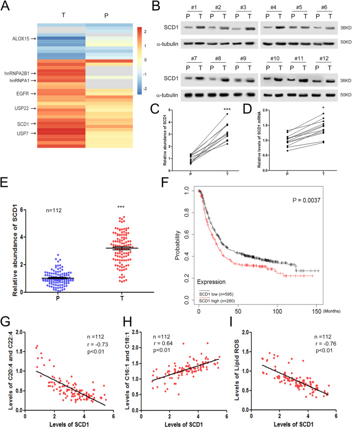
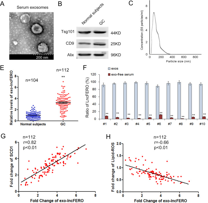
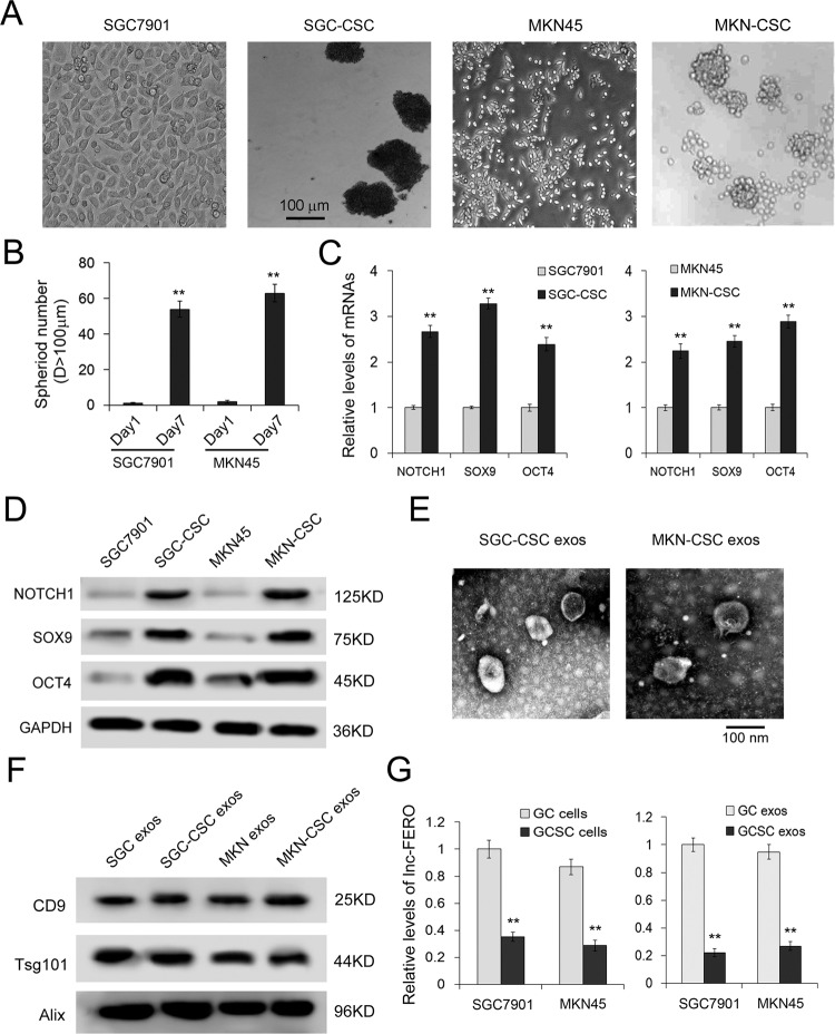
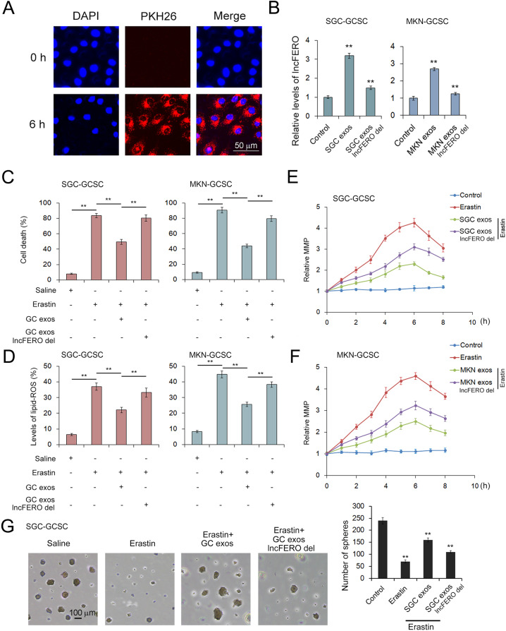
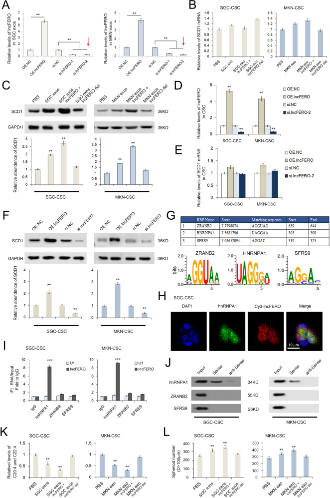
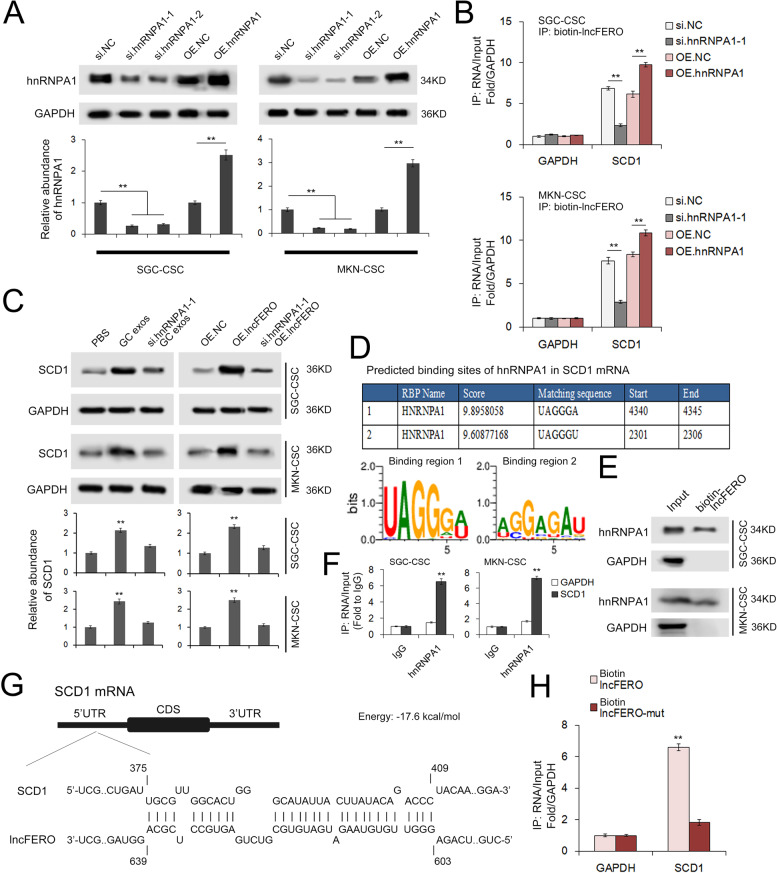
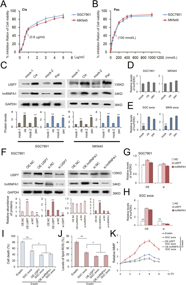
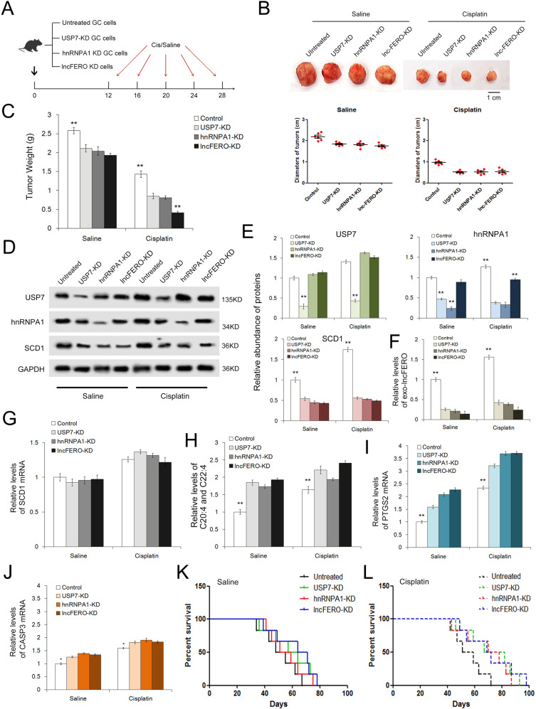
Cancer stem cells (CSCs) are an important cause of tumor recurrence and drug resistance. As a new type of cell death that relies on iron ions and is strictly regulated by intracellular and extracellular signals, the role of ferroptosis in tumor stem cells deserves extensive attention. Mass spectrum was applied to screen for ferroptosis-related proteins in gastric cancer (GC). Sphere-formation assay was used to estimate the stemness of gastric cancer stem cells (GCSCs). Exosomal lnc-ENDOG-1:1 (lncFERO) was isolated by ultracentrifugation. Ferroptosis was induced by erastin and was assessed by detecting lipid ROS, mitochondrial membrane potential, and cell death. Furthermore, a series of functional in vitro and in vivo experiments were conducted to evaluate the effects of lncFERO on regulating ferroptosis and chemosensitivity in GCSCs. Here, we showed that stearoyl-CoA-desaturase (SCD1) played a key role in regulating lipid metabolism and ferroptosis in GCSCs. Importantly, exosomal lncFERO (exo-lncFERO) derived from GC cells was demonstrated to promote SCD1 expression by directly interacting with SCD1 mRNA and recruiting heterogeneous nuclear ribonucleoprotein A1 (hnRNPA1), which resulted in the dysregulation of PUFA levels and the suppression of ferroptosis in GCSCs. Moreover, we found that hnRNPA1 was also involved in lncFERO packing into exosomes in GC cells, and both in vitro and in vivo data suggested that chemotoxicity induced lncFERO secretion from GC cells by upregulating hnRNPA1 expression, leading to enhanced stemness and acquired chemo-resistance. All these data suggest that GC cells derived exo-lncFERO controls GCSC tumorigenic properties through suppressing ferroptosis, and targeting exo-lncFERO/hnRNPA1/SCD1 axis combined with chemotherapy could be a promising CSC-based strategy for the treatment of GC.


Conflict of interest statement

The authors declare no competing interests.

Figures

Fig. 1
Fig. 1. SCD1 is related to lipid metabolism and ferroptosis in gastric cancer.
A Mass spectrum analysis of protein expression changes in gastric tumor tissues (n = 45). The heatmap depicts the relative protein abundance in tumor tissues (T) and paired paracarcinoma tissues (P). B Validation of SCD1 dysregulation in GC by using western blotting analysis (n = 12). C Quantitative analysis of (B) (n = 12). D Relative levels of SCD1 mRNA in gastric tumor tissues (n = 12). E Validation of SCD1 dysregulation in GC tissues using protein microarray (n = 112). F High expression of SCD1 predicts poor overall survival in GC. Patients were divided into the SCD1 high group (n = 280) and SCD1 low group (n = 595) based on the average value of SCD1 mRNA levels. G SCD1 is negatively correlated with polyunsaturated fatty acids (PUFAs) (C20:4 and C22:4) (n = 112). H SCD1 is positively correlated with monounsaturated fatty acids (MUFAs) (C16:1 and C18:1) (n = 112). I SCD1 is negatively correlated with lipid ROS production (n = 112). *p < 0.05 and ***p < 0.001.
Fig. 2
Fig. 2. Exosomal lncFERO is positively linked with SCD1 expression.
A Serum exosomes were observed by using an electron microscope (scale bar, 200 nm). B Detection of exosome markers, Tsg101, CD9, and Alix, by western blotting assay. C Nanotracking analysis of the diameters of vesicles. E Relative levels of exo-lncFERO in normal subjects (n = 104) and GC patients (n = 112). F Relative levels of exo-lncFERO in the exos and exo-free serum (n = 10). G Exo-lncFERO is positively correlated with SCD1 (n = 112). H Exo-lncFERO is negatively correlated with lipid ROS levels (n = 112). **p < 0.01.
Fig. 3
Fig. 3. The different expression characters of lncFERO in GC cells and GC stem cells.
A Sphere-formation assays were performed in indicated cell lines. B Number of tumorspheres (n = 3). C qRT-PCR analysis of stemness-associated genes, including NOTCH1, SOX9, and OCT4, in GC and GCSC cell lines (n = 3). D Detection of stemness-associated genes by western blotting assay (n = 3). E Exosomes images obtained from GCSCs were observed by using an electron microscope (scale bar, 100 nm). F Detection of exosome markers, CD9, Tsg101, and Alix, by western blotting assay. G Relative levels of lncFERO in GC cells and GCSCs (n = 3). **p < 0.01.
Fig. 4
Fig. 4. Exosome-delivered lncFERO suppresses ferroptosis in GCSCs.
A Confocal microscopy image of the internalization of fluorescently labeled GC exosomes in GCSCs. B Effects of GC exosomes on lncFERO levels in GCSCs (n = 3). CF Exo-lncFERO derived from GC cells suppresses erastin-induced ferroptosis in GCSCs. GC exo-lncFERO decreases erastin-induced cell death (C) (n = 3), inhibits lipid ROS accumulation (D) (n = 3), and reduces abnormal increases in MMP (E, F) (n = 3). G Effects of erastin and exo-lnc-FERO on sphere formation of GCSCs (n = 3). **p < 0.01.
Fig. 5
Fig. 5. LncFERO promotes SCD1 expression by recruiting hnRNPA1 in GCSCs.
A Levels of lncFERO in exosomes isolated from GC cells treated with lncFERO-OE plasmids or siRNAs (n = 3). B Effects of GC-secreted lncFERO on SCD1 mRNA levels in GCSCs (n = 3). C Effects of GC-secreted lncFERO on SCD1 expression in GCSCs (n = 3). D lncFERO in GCSCs was overexpressed or knocked down (n = 3). E Effects of lncFERO on the expression of SCD1 mRNA in GCSCs (n = 3). F Western blotting analysis of SCD1 expression in GCSCs with overexpression or knockdown of lncFERO (n = 3). G RBPDB analysis of the specific interaction between lncFERO and RBP motifs. H The colocalization of Cy3-lncFERO and hnRNPA1 in GCSCs (n = 3). I RNA immunoprecipitation assay shows the direct interaction between lncFERO and hnRNPA1 in GCSCs (n = 3). J Detection of hnRNPA1 protein in the samples derived from lncFERO pulldowns performed in GCSCs. K GC exosomal lncFERO decreased PUFA levels in GCSCs (n = 3). L GC exosomal lncFERO promoted the stemness of GCSCs (n = 3). **p < 0.01 and ***p < 0.001.
Fig. 6
Fig. 6. LncFERO interacts with SCD1 mRNA and promotes SCD1 translation by recruiting hnRNPA1.
A hnRNPA1 in GCSCs was overexpressed or knocked down (n = 3). B hnRNPA1 stabilizes the interaction between lncFERO and SCD1 mRNA (n = 3). C GC exosomal lncFERO depends on hnRNPA1 to promote SCD1 expression in GCSCs (n = 3). D RBPDB analysis of the specific interaction between hnRNPA1 and SCD1. E Detection of hnRNPA1 protein in the samples derived from lncFERO pulldowns performed in GCSCs (n = 3). F RIP shows the direct interaction between SCD1 mRNA and hnRNPA1 in GCSCs (n = 3). G Predicted binding sites of lncFERO in the 5’UTR of SCD1 mRNA. H Capture of SCD1 mRNA by biotin-labeled lncFERO (n = 3). **p < 0.01.
Fig. 7
Fig. 7. Chemotoxicity promotes lncFERO secretion from GC cells via the USP7/hnRNPA1 axis.
A, B The effects of gradient doses of cisplatin (A) and paclitaxel (B) on the viability of SGC7901 and MKN45 cells (n = 3). C The expression of USP7 and hnRNPA1 in GC cells treated with sublethal doses of cisplatin (0.8 μg/mL) and paclitaxel (100 nmol/L) (n = 3). D Effects of cisplatin and paclitaxel on the expression of lncFERO in GC cells (n = 3). E Effects of cisplatin and paclitaxel on the secretion of lncFERO in GC exos (n = 3). F WB analysis of USP7 and hnRNPA1 in SGC7901 cells treated with corresponding overexpression plasmids or siRNAs (n = 3). G Effects of USP7 and hnRNPA1 on the expression of lncFERO in SGC7901 cells (n = 3). H Effects of USP7 and hnRNPA1 on the expression of lncFERO in SGC exos (n = 3). IK Exosomes derived from SGC7901 cells overexpressing USP7 and hnRNPA1 suppressed erastin-induced cell death (I), lipid ROS production (J), and the MMP increase (K) in GCSCs (n = 3). *p < 0.05 and **p < 0.01.
Fig. 8
Fig. 8. In vivo role of the USP7/hnRNPA1/lncFERO pathway in regulating ferroptosis and chemosensitivity of gastric tumors.
A Schematic description of the experimental design used to establish the animal model. B Images and the diameters of tumors in each group (n = 6). C Weight measurements of the tumors described above (n = 6). D WB analysis of USP7, hnRNPA1 and SCD1 in tumor tissues (n = 3). E Quantitative analysis of (D) (n = 3). F Relative levels of serum exo-lncFERO in each group (n = 6). G Relative levels of SCD1 mRNA in tumor tissues (n = 6). H Relative levels of PUFAs in tumor tissues (n = 6). I, J Quantification of a ferroptosis marker (I) and an apoptosis marker (J) in each group (n = 6). K, L Kaplan–Meier curves of mice in the saline-treated groups (K) and cisplatin-treated groups (L) (n = 6). **p < 0.01.

References

    1. Jung E, Osswald M, Ratliff M, Dogan H, Xie R, Weil S, et al. Tumor cell plasticity, heterogeneity, and resistance in crucial microenvironmental niches in glioma. Nat Commun. 2021;12:1014. - PMC - PubMed
    1. Karthikeyan S, Waters IG, Dennison L, Chu D, Donaldson J, Shin DH, et al. Hierarchical tumor heterogeneity mediated by cell contact between distinct genetic subclones. J Clin Invest. 2021;131:e143557.. - PMC - PubMed
    1. Konen JM, Rodriguez BL, Padhye A, Ochieng JK, Gibson L, Diao L, et al. Dual inhibition of MEK and AXL targets tumor cell heterogeneity and prevents resistant outgrowth mediated by the epithelial-to-mesenchymal transition in NSCLC. Cancer Res. 2021;81:1398–412. - PMC - PubMed
    1. Giraud J, Molina-Castro S, Seeneevassen L, Sifré E, Izotte J, Tiffon C, et al. Verteporfin targeting YAP1/TAZ-TEAD transcriptional activity inhibits the tumorigenic properties of gastric cancer stem cells. Int J Cancer. 2020;146:2255–67. - PubMed
    1. Yang L, Shi P, Zhao G, Xu J, Peng W, Zhang J, et al. Targeting cancer stem cell pathways for cancer therapy. Signal Transduct Target Ther. 2020;5:8. - PMC - PubMed

Publication types

MeSH terms