. 2021 Nov 29;11(1):23094.
doi: 10.1038/s41598-021-02589-1.

Photocrosslinked gelatin hydrogel improves wound healing and skin flap survival by the sustained release of basic fibroblast growth factor

Affiliations

Photocrosslinked gelatin hydrogel improves wound healing and skin flap survival by the sustained release of basic fibroblast growth factor

Toshihiro Kushibiki et al. Sci Rep. .

Abstract

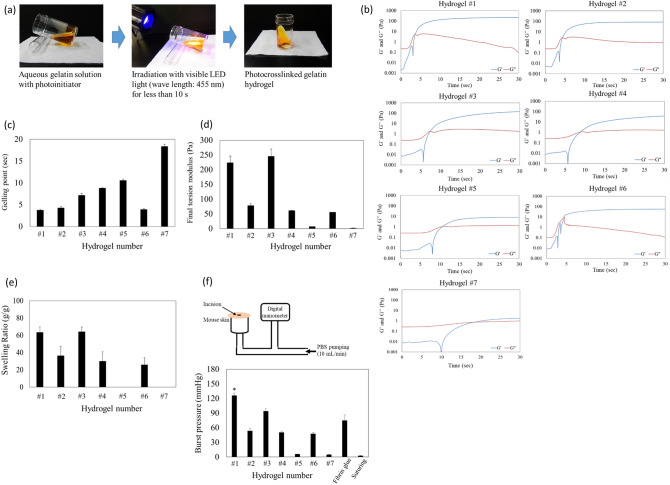
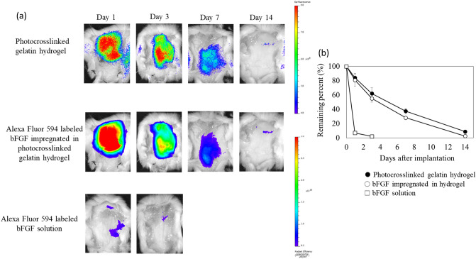
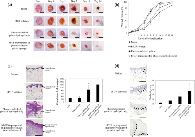
Biomaterials traditionally used for wound healing can act as a temporary barrier to halt bleeding, prevent infection, and enhance regeneration. Hydrogels are among the best candidates for wound healing owing to their moisture retention and drug-releasing properties. Photo-polymerization using visible light irradiation is a promising method for hydrogel preparation since it can easily control spatiotemporal reaction kinetics and rapidly induce a single-step reaction under mild conditions. In this study, photocrosslinked gelatin hydrogels were imparted with properties namely fast wound adherence, strong wet tissue surface adhesion, greater biocompatibility, long-term bFGF release, and importantly, ease of use through the modification and combination of natural bio-macromolecules. The production of a gelatin hydrogel made of natural gelatin (which is superior to chemically modified gelatin), crosslinked by visible light, which is more desirable than UV light irradiation, will enable its prolonged application to uneven wound surfaces. This is due to its flexible shape, along with the administration of cell growth factors, such as bFGF, for tissue regeneration. Further, the sustained release of bFGF enhances wound healing and skin flap survival. The photocrosslinking gelatin hydrogel designed in this study is a potential candidate to enhance wound healing and better skin flap survival.


Conflict of interest statement

The authors declare no competing interests.

Figures

Figure 1
Figure 1
Formation and characterization of hydrogel crosslinked by light irradiation. (a) Process of hydrogel formation by photocrosslinking. (be) Mechanical properties of the photocrosslinked gelatin hydrogels. (b) To monitor the gelling process, a dynamic time-sweep rheological analysis was conducted using an in situ photo-rheometer (HAAKE Mars and LED 455 nm) showing the formation kinetics for photocrosslinked gelatin hydrogels. (c) Gelling points and (d) the final torsion moduli, G′, of different hydrogels. The exposure time for all gelling measurements was 120 s (error bars, mean ± SD; n = 3 per group). (e) Swelling ratios of different hydrogels (n = 8 per group) after 24 h incubation in PBS at 37 °C (error bars, mean ± SD). (f) Burst pressure of photocrosslinked gelatin hydrogels and fibrin glues. Schematic illustration of the experimental procedure and pressure chamber for burst adhesion testing using punctured and then sealed mouse skin tissue. The observed burst pressure of photocrosslinked gelatin hydrogels and fibrin glue were measured (mean ± SD; *p < 0.005 against fibrin glue; n = 5 per group).
Figure 2
Figure 2
Evaluation of in vitro and in vivo biocompatibility of photocrosslinked gelatin hydrogels. (a) Cytotoxicity towards L929 fibroblasts after incubation with hydrogel extracts for 1, 3, 5, and 7 days (n = 8 per group). Experimental data were represented by dividing each value by that of the control group (incubated with normal culture medium) for each day (error bars, mean ± SD). (b) Live/dead staining of L929 fibroblasts encapsulated in the hydrogels after 3 and 5 days of incubation. L929 fibroblasts were stained with calcein-AM to detect living cells (green) and ethidium homodimer-1 to detect dead cells (red). Scale bar: 300 μm. (c) Macroscopic appearance of excised photocrosslinked gelatin hydrogel #1 implants after 1, 3, 7, and 14 days of implantation (n = 5 per group). At designated time intervals, the fluorescence of the remaining hydrogel in mouse subcutaneous tissue was imaged using the IVIS Lumina XR, and the fluorescence intensity was analyzed with Living Image Software (PerkinElmer Inc., MA). Scale bar: 10 mm. (d) After imaging, the subcutaneous tissues were processed for histological analyses. Pathological staining with hematoxylin and eosin showed several inflammatory cells near the hydrogel–tissue interface on day 1, indicating a post-operative inflammatory response. However, this inflammatory response disappeared within 3 days. The yellow arrowhead indicates the remaining hydrogel. Scale bar: 200 μm.
Figure 3
Figure 3
In vivo release of bFGF from photocrosslinked gelatin hydrogel. (a) Decreasing patterns of bFGF impregnated in photocrosslinked gelatin hydrogel after subcutaneous implantation into mice. The residual bFGF in the photocrosslinked gelatin hydrogels decreased with implantation time. In contrast, the aqueous solution of bFGF (without gelatin hydrogel) rapidly disappeared from the injected site within 3 days. (b) The time profile of bFGF retention was in good accordance with that of hydrogel degradation. These findings indicate the possibility that bFGF is released from the photocrosslinked gelatin hydrogel in the body due to hydrogel biodegradation.
Figure 4
Figure 4
Time course of wound closure and histological evaluation in diabetic mice after sustained release of bFGF from photocrosslinked gelatin hydrogel. (a) Images of in vivo wound closure studies for saline, bFGF solution (Fiblast spray), and photocrosslinked gelatin hydrogels with and without bFGF. Macroscopic photographs of wounds at days 5, 7, and 10 demonstrated significantly faster wound closure in the group treated with photocrosslinked gelatin hydrogels with bFGF. Scale bar: 5 mm. (b) Average wound closure from days 0 to 14 [expressed as a percent of the day 0 control (100%)]. On day 7, the group treated photocrosslinked gelatin hydrogels with bFGF showed a greater extent of wound closure when compared to that in saline and bFGF solution groups. *p < 0.005 vs saline group. (c) Histology of wounds treated with saline, bFGF solution (Fiblast spray), and photocrosslinked gelatin hydrogels with and without bFGF on post-operative day 7. The surfaces of the wounds treated with the photocrosslinked gelatin hydrogels with bFGF showed epithelium neoformation. This wound also showed the formation of granulation tissue with the highest thickness. The increased number of fibroblasts and the collagen amount in the wounds treated with bFGF-containing photocrosslinked gelatin hydrogels significantly contributed to enhanced granulation tissue formation. Scale bar: 300 μm; *p < 0.005. (d) Histological evaluation of the blood capillaries. The wounds treated with photocrosslinked gelatin hydrogels with bFGF had an increased distribution of blood capillaries (arrowhead) compared to that with saline and bFGF solution (Fiblast spray) on post-operative day 7. Scale bar: 300 μm; *p < 0.005 vs saline group.
Figure 5
Figure 5
Improved flap survival in mice by sustained release of bFGF from photocrosslinked gelatin hydrogel. (a) Comparison of survival of random pattern skin flaps among saline, bFGF solution (Fiblast spray), and photocrosslinked gelatin hydrogels with and without bFGF on post-operative day 10. Assessment of tissue viability at the random skin flap area in both groups of transgenic mice expressing FRET-based biosensors, named “ATeam,” for visualization of ATP levels in living cells. Representative images showing the ATP level in the skin flap was greater in the photocrosslinked gelatin hydrogel with bFGF group on post-operative day 10 than in the other groups. Scale bar: 10 mm. The color bar shows the FRET ratio. The higher the FRET ratio, the higher the intracellular ATP content, as detected using ATeam. (b) Histological evaluation of the blood capillaries. Flaps treated with bFGF-containing photocrosslinked gelatin hydrogels had an increased distribution of blood capillaries compared to that with saline and bFGF solution (Fiblast spray) on post-operative day 10. Scale bar: 100 μm; *p < 0.005 vs saline group.

References

    1. Guo S, Dipietro LA. Factors affecting wound healing. J. Dent. Res. 2010;89:219–229. doi: 10.1177/0022034509359125. - DOI - PMC - PubMed
    1. Maalej H, et al. Rhelogical, dermal wound healing and in vitro antioxidant properties of exopolysaccharide hydrogel from Pseudomonas stutzeri AS22. Colloids Surf. B Biointerfaces. 2014;123:814–824. doi: 10.1016/j.colsurfb.2014.10.017. - DOI - PubMed
    1. Lopez AD, Mathers CD, Ezzati M, Jamison DT, Murray CJ. Global and regional burden of disease and risk factors, 2001: Systematic analysis of population health data. Lancet. 2006;367:1747–1757. doi: 10.1016/s0140-6736(06)68770-9. - DOI - PubMed
    1. Wu J, Zhao X, Wu D, Chu CC. Development of a biocompatible and biodegradable hybrid hydrogel platform for sustained release of ionic drugs. J. Mater. Chem. B. 2014;2:6660–6668. doi: 10.1039/c4tb00576g. - DOI - PubMed
    1. Yadav S, Sharma AK, Kumar P. Nanoscale self-assembly for therapeutic delivery. Front. Bioeng. Biotechnol. 2020;8:127. doi: 10.3389/fbioe.2020.00127. - DOI - PMC - PubMed

MeSH terms