. 2021 Nov 29;11(1):23086.
doi: 10.1038/s41598-021-02550-2.

The implications of hyperoxia, type 1 diabetes and sex on cardiovascular physiology in mice

Affiliations

The implications of hyperoxia, type 1 diabetes and sex on cardiovascular physiology in mice

Katarina Bojkovic et al. Sci Rep. .

Abstract

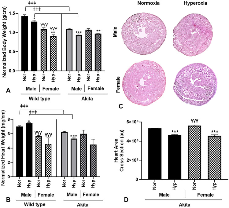
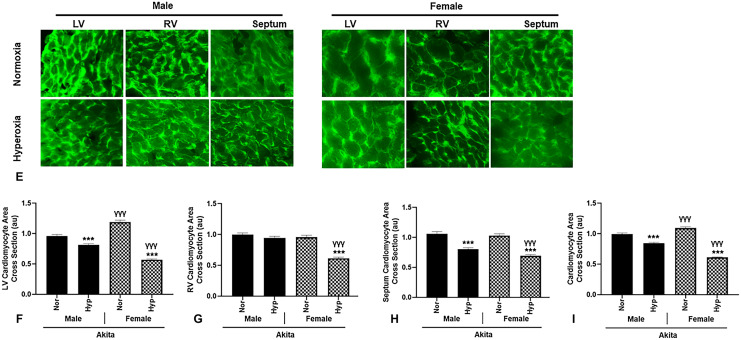
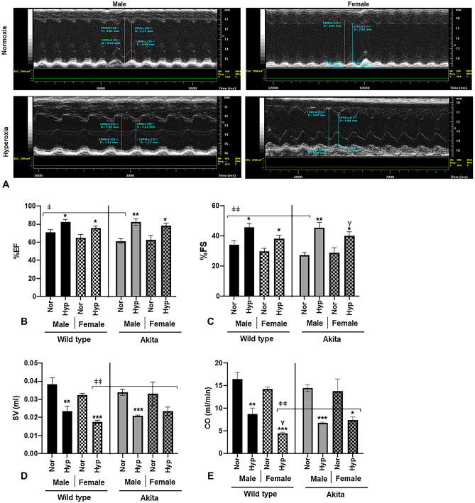
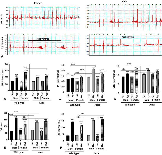
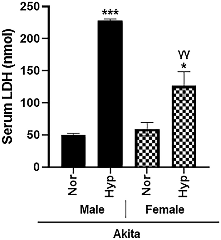
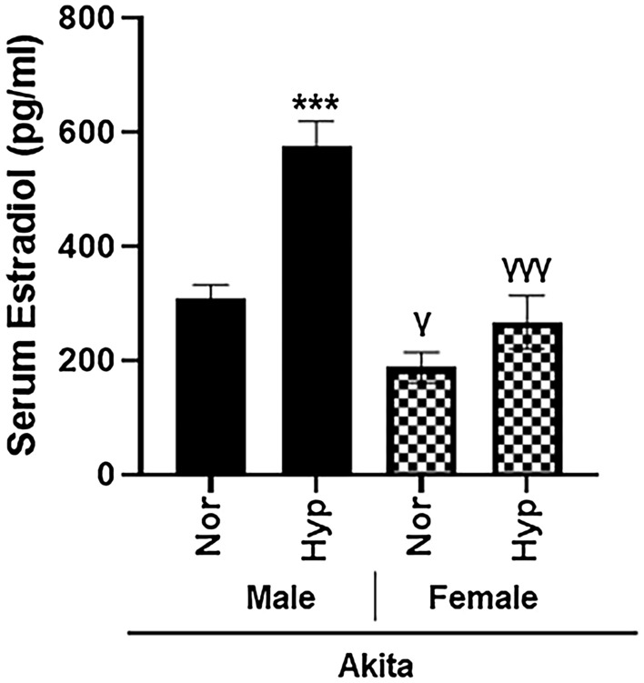
Oxygen supplementation, although a cornerstone of emergency and cardiovascular medicine, often results in hyperoxia, a condition characterized by excessive tissue oxygen which results in adverse cardiac remodeling and subsequent injurious effects to physiological function. Cardiac remodeling is further influenced by various risk factors, including pre-existing conditions and sex. Thus, the purpose of this experiment was to investigate cardiac remodeling in Type I Diabetic (Akita) mice subjected to hyperoxic treatment. Overall, we demonstrated that Akita mice experience distinct challenges from wild type (WT) mice. Specifically, Akita males at both normoxia and hyperoxia showed significant decreases in body and heart weights, prolonged PR, QRS, and QTc intervals, and reduced %EF and %FS at normoxia compared to WT controls. Moreover, Akita males largely resemble female mice (both WT and Akita) with regards to the parameters studied. Finally, statistical analysis revealed hyperoxia to have the greatest influence on cardiac pathophysiology, followed by sex, and finally genotype. Taken together, our data suggest that Type I diabetic patients may have distinct cardiac pathophysiology under hyperoxia compared to uncomplicated patients, with males being at high risk. These findings can be used to enhance provision of care in ICU patients with Type I diabetes as a comorbid condition.


Conflict of interest statement

The authors declare no competing interests.

Figures

Figure 1
Figure 1
Akita male mice at both normoxia and hyperoxia experience a significant decrease in body and heart weight. (A) Body weight normalized to tibia length (g/cm) for all experimental groups, (B) Heart weight normalized to tibia length for all experimental groups (mg/cm), (C) H&E histological cross sections of Akita male and female mice under normoxia and hyperoxia, (D) Heart area cross sections (au), (E) WGA stained cardiac myocytes stained from Akita male and female mice. (FI) Cardiac myocyte area of Akita male and female mice under normoxia or hyperoxia shown as follows: (F) LV cardiomyocte area, (G) RV cardiomyocte area, (H) Septum cardiomyocte area, (I) Pooled cardiomyocte area. Error bars represent ± SEM. *p < 0.05, **p < 0.005 ***p < 0.0005. *compares effects of hyperoxia and normoxia of same gender and strain; Y compares male and female mice of the same strain and treatment; ‡compares WT and Akita of same gender and treatment.
Figure 1
Figure 1
Akita male mice at both normoxia and hyperoxia experience a significant decrease in body and heart weight. (A) Body weight normalized to tibia length (g/cm) for all experimental groups, (B) Heart weight normalized to tibia length for all experimental groups (mg/cm), (C) H&E histological cross sections of Akita male and female mice under normoxia and hyperoxia, (D) Heart area cross sections (au), (E) WGA stained cardiac myocytes stained from Akita male and female mice. (FI) Cardiac myocyte area of Akita male and female mice under normoxia or hyperoxia shown as follows: (F) LV cardiomyocte area, (G) RV cardiomyocte area, (H) Septum cardiomyocte area, (I) Pooled cardiomyocte area. Error bars represent ± SEM. *p < 0.05, **p < 0.005 ***p < 0.0005. *compares effects of hyperoxia and normoxia of same gender and strain; Y compares male and female mice of the same strain and treatment; ‡compares WT and Akita of same gender and treatment.
Figure 2
Figure 2
Akita mice display distinct functional parameters compared to WT mice (A) percent ejection fraction (FS), (B) percent fractional shortening (EF), (C) stroke volume (SV), and (D) cardiac output (CO) in hyperoxia/normoxia treated Akita and wild type mice, (E) Optical traces for male and female Akita mice in normoxic and hyperoxic conditions. Error bars represent ± SEM. *p < 0.05, **p < 0.005 ***p < 0.0005. *compares effects of hyperoxia and normoxia of same gender and strain; Y compares male and female mice of the same strain and treatment; ‡compares WT and Akita of same gender and treatment.
Figure 3
Figure 3
Hyperoxia induces bradyarrhythmias in Akita (and WT) mice (A) Original ECG traces in lead II mode in normoxia/hyperoxia treated Akita male and female mice, (B) RR interval, (C) PR interval, (D) QRS interval, (E) QTc interval, (F) JT interval. Error bars represent ± SEM. *p < 0.05, **p < 0.005 ***p < 0.0005. *compares effects of hyperoxia and normoxia of same gender and strain; Y compares male and female mice of the same strain and treatment; ‡compares WT and Akita of same gender and treatment.
Figure 4
Figure 4
Hyperoxia induce cardiotoxicity in Akita mice as evident by elevated levels of serum LDH in both sexes. Lactic Dehydrogenase (LDH) measured in serum from normoxia and hyperoxia WT and Akita groups. Error bars represent ± SEM. *p < 0.05, **p < 0.005 ***p < 0.0005. *compares effects of hyperoxia and normoxia of same gender and strain; Y compares male and female mice of the same strain and treatment.
Figure 5
Figure 5
Dysregulation of serum estradiol in Akita male mice in both normal air and hyperoxia. Estradiol levels were measured in serum from normoxia and hyperoxia WT and Akita groups using ELISA kit. Error bars represent ± SEM. *p < 0.05, **p < 0.005 ***p < 0.0005. *compares effects of hyperoxia and normoxia of same gender and strain; Y compares male and female mice of the same strain and treatment.

Similar articles

Cited by

References

    1. Bitterman, H. Bench-to-bedside review: Oxygen as a drug. Crit. Care13, 205. 10.1186/cc7151 (2009). - PMC - PubMed
    1. Ni, Y.-N., Wang, Y.-M., Liang, B.-M. & Liang, Z.-A. The effect of hyperoxia on mortality in critically ill patients: A systematic review and meta analysis. BMC Pulm. Med.19, 53. 10.1186/s12890-019-0810-1 (2019). - PMC - PubMed
    1. Helmerhorst, H. J., Schultz, M. J., van der Voort, P. H., de Jonge, E. & van Westerloo, D. J. Bench-to-bedside review: The effects of hyperoxia during critical illness. Crit Care19, 284. 10.1186/s13054-015-0996-4 (2015). - PMC - PubMed
    1. Hanidziar, D. & Robson, S. C. Hyperoxia and modulation of pulmonary vascular and immune responses in COVID-19. Am. J. Physiol. Lung Cell. Mol. Physiol.320, L12–L16. 10.1152/ajplung.00304.2020 (2021). - PMC - PubMed
    1. Wunsch, H. et al. ICU occupancy and mechanical ventilator use in the United States. Crit. Care Med.41, 2712–2719. 10.1097/CCM.0b013e318298a139 (2013). - PMC - PubMed

Publication types