[Preprint]. 2021 Nov 22:2021.11.19.469183.
doi: 10.1101/2021.11.19.469183.

Self-assembling short immunostimulatory duplex RNAs with broad spectrum antiviral activity

Affiliations

Self-assembling short immunostimulatory duplex RNAs with broad spectrum antiviral activity

Longlong Si et al. bioRxiv. .

Update in

  • Self-assembling short immunostimulatory duplex RNAs with broad-spectrum antiviral activity.
    Si L, Bai H, Oh CY, Jiang A, Hong F, Zhang T, Ye Y, Jordan TX, Logue J, McGrath M, Belgur C, Calderon K, Nurani A, Cao W, Carlson KE, Prantil-Baun R, Gygi SP, Yang D, Jonsson CB, tenOever BR, Frieman M, Ingber DE. Si L, et al. Mol Ther Nucleic Acids. 2022 Sep 13;29:923-940. doi: 10.1016/j.omtn.2022.08.031. Epub 2022 Aug 24. Mol Ther Nucleic Acids. 2022. PMID: 36032397 Free PMC article.

Abstract

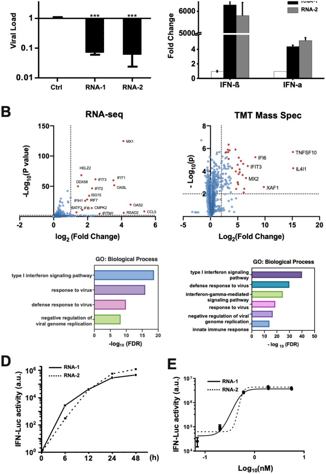
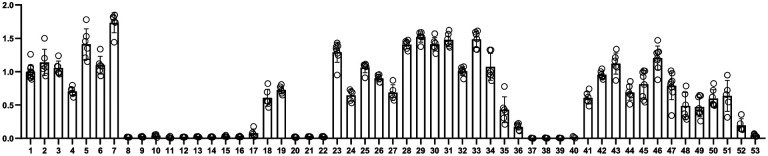
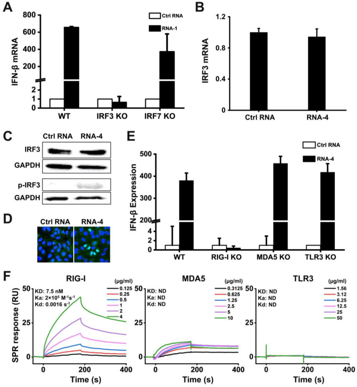
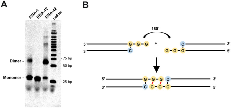
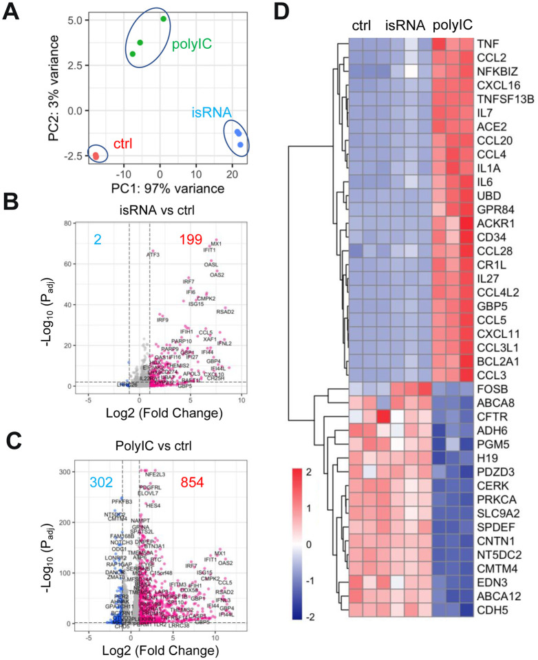
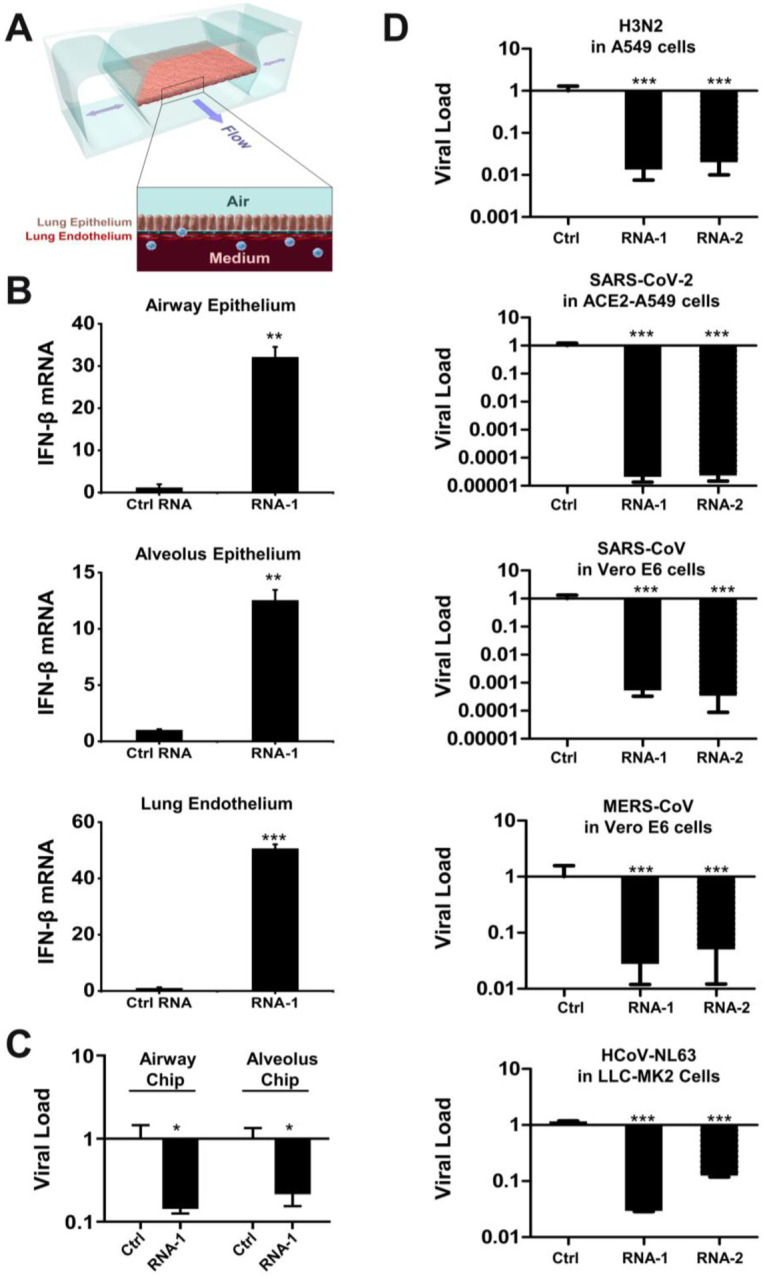
The current COVID-19 pandemic highlights the need for broad-spectrum antiviral therapeutics. Here we describe a new class of self-assembling immunostimulatory short duplex RNAs that potently induce production of type I and type III interferon (IFN-I and IFN-III), in a wide range of human cell types. These RNAs require a minimum of 20 base pairs, lack any sequence or structural characteristics of known immunostimulatory RNAs, and instead require a unique conserved sequence motif (sense strand: 5'-C, antisense strand: 3'-GGG) that mediates end-to-end dimer self-assembly of these RNAs by Hoogsteen G-G base-pairing. The presence of terminal hydroxyl or monophosphate groups, blunt or overhanging ends, or terminal RNA or DNA bases did not affect their ability to induce IFN. Unlike previously described immunostimulatory siRNAs, their activity is independent of TLR7/8, but requires the RIG-I/IRF3 pathway that induces a more restricted antiviral response with a lower proinflammatory signature compared with poly(I:C). Immune stimulation mediated by these duplex RNAs results in broad spectrum inhibition of infections by many respiratory viruses with pandemic potential, including SARS-CoV-2, SARS-CoV, MERS-CoV, and influenza A, as well as the common cold virus HCoV-NL63 in both cell lines and human Lung Chips that mimic organ-level lung pathophysiology. These short dsRNAs can be manufactured easily, and thus potentially could be harnessed to produce broad-spectrum antiviral therapeutics at low cost.


Conflict of interest statement

Conflict of Interest

D.E.I. is a founder, board member, SAB chair, and equity holder in Emulate Inc. D.E.I., L. S., H. B., C.O., and R.P. are inventors on relevant patent applications held by Harvard University.

Figures

Figure 1.
Figure 1.. Discovery of new immunostimulatory RNAs.
(A) A549 cells were transfected with RNA-1, RNA-2, or a scrambled duplex RNA control, and infected with influenza A/WSN/33 (H1N1) virus (MOI=0.01) 24 hours later. Titers of progeny viruses in medium supernatants collected at 48 h post-infection were determined by quantifying plaque forming units (PFUs); data are shown as % viral infection measured in the cells treated with the control RNA (Data shown are mean ± standard deviation; N =3; ***, p < 0.001). (B) A549 cells were transfected with RNA-1 or a scrambled dsRNA control, collected at 48 h, and analyzed by RNA-seq (left) or TMT Mass Spec (right). Differentially expressed genes (DEGs) from RNA-seq or proteins from TMT Mass Spec are shown in volcano plots (top) and results of GO Enrichment analysis performed for the DEGs are shown at the bottom (N = 3). (C) qPCR analysis of cellular IFN-β and IFN-α RNA levels at 48 h after A549 cells were transfected with RNA-1, RNA-2, or scrambled dsRNA control (N = 3). (D) RNA-mediated production kinetics of IFN production in wild-type A549-Dual cells that were transfected with RNA-1, RNA-2, or scramble RNA control measured using a Quanti-Luc assay. OD values from cells transfected with the scrambled RNA control were subtracted as background (N = 6). (E) Dose-dependent induction of IFN by RNA-1 and −2 in A549-Dual cells compared to scrambled RNA control measured at 48 h post-transfection (control OD values were subtracted as background; N = 6).
Figure 2.
Figure 2.. Comparison of the immunostimulatory activities of different RNAs.
A549-Dual cells were transfected with indicated duplex RNAs for 48 h, and then activation of the IFN pathway was measured by quantifying luciferase reporter activity. The immunostimulatory activity of RNA-1 was set as 1 (N = 6).
Figure 3.
Figure 3.. Immunostimulatory RNAs induce IFN-I production through RIG-I-IRF3 pathway.
(A) Wild-type (WT) HAP1 cells, IRF3 knockout HAP1 cells, or IRF7 knockout HAP1 cells were transfected with RNA-1 or scrambled RNA control for 48 h, and IFN-β mRNA levels were quantified by qPCR. Data are shown as fold change relative to the scrambled RNA control (N = 3). Note that IRF3 knockdown completely abolished the IFN-β response. (B) IRF3 mRNA levels measured in A549 cells transfected with immunostimulatory RNA-4 or a scrambled RNA control, as determined by qPCR and 48 h post-transfection (data are shown as fold change relative to the control RNA; N = 3). (C) Total IRF3 protein and phosphorylated IRF3 detected in A549 cells transfected with RNA-4 or scrambled RNA control at 48 h post transfection as detected by Western blot analysis (GAPDH was used as a loading control). (D) Immunofluorescence micrographs showing the distribution of phosphorylated IRF3 in A549 cells transfected with RNA-4 or scrambled RNA control at 48 h post transfection (Green, phosphorylated IRF3; blue, DAPI-stained nuclei; arrowheads, nuclei expressing phosphorylated IRF3). (E) Wild-type (WT) A549-Dual cells, RIG-I knockout A549-Dual cells, MDA5 knockout A549-Dual cells, or TLR3 knockout A549 cells were transfected with immunostimulatory RNA-4 or a scrambles RNA control and 48 h later, IFN-β expression levels were quantified using the Quanti-Luc assay or qPCR (data are shown as fold change relative to the scrambled RNA control; N = 6). Note that RIG-I knockout abolished the ability of the immunostimulatory RNAs to induce IFN-β. (F) SPR characterization of the binding affinity between cellular RNA sensors (RIG-I, MDA5, and TLR3) and RNA-1, which were immobilized on a streptavidin (SA) sensor chip. Equilibrium dissociation constant (KD), association rate constant (Ka), and dissociation rate constant (Kd) are labeled on the graphs.
Figure 4.
Figure 4.. The common motif mediates the formation of duplex RNA dimers via intramolecular G-quadruplex formed by GG overhang.
(A) The image of native gel electrophoresis showing the formation of RNA-1 dimer. 1 uL of 10 uM RNA samples were loaded. RNA-12 and RNA-42 were used as negative and positive control, respectively. (B) The diagram showing the structure of ‘end-to-end’ RNA-1 dimer due to terminal G-G Hoogsteen paring.
Figure 5.
Figure 5.. Immunostimulatory RNAs elicit responses with a stronger antiviral component and a lower proinflammatory component.
(A) Principal component analysis of A549 cells transcriptomes when transfected with scrambled dsRNA (ctrl), RNA-1 (isRNA) or poly(I:C) for 48 hours. N=3. (B and C) Volcano plots showing significant upregulated genes (red) or downregulated genes (blue) in isRNA transfected (B) or poly(I:C) transfected (C) A549 cells. Threshold for fold change = 2, threshold for Padj = 0.01. (D). Heat map showing top upregulated inflammatory genes and top downregulated genes involved in ion transport and cell-cell adhesion in the poly(I:C) transfected but not in the isRNA transfected A549 cells.
Figure 6.
Figure 6.. Immunostimulatory RNAs induce IFN-β production in differentiated human lung epithelial and endothelial cells in Organ Chips and exhibit broad spectrum inhibition of infection by H3N2 influenza virus, SARS-CoV-2, SARS-CoV-1, MERS-CoV, and HCoV-NL63.
(A) Schematic diagram of a cross-section through the human Lung-on-Chip, which faithfully recapitulate human lung physiology and pathophysiology. (B) Human Lung Airway and Alveolus Chips were transfected with RNA-1 or scrambled RNA control by perfusion through both channels of the chip and 48 h later, the epithelial and endothelial cells were collected for detection of IFN-β mRNA by qPCR (data are presented as fold change relative to the RNA control; N = 3; *, p < 0.05; ***, p < 0.001). (C) Effects of treatment with RNA-1 or a scrambled control in the human Lung Airway Chips or human Lung Alveolus Chips infected with influenza A/HK/8/68 (H3N2) (MOI = 0.1) at 24 h after RNA-1 treatment. Viral load was determined by quantifying the viral NP gene by qPCR in cell lysates at 48 h after infection. Results are shown as fold change relative to RNA control; N=3; *, p < 0.05. (D) Treatment with immunostimulatory duplex RNAs resulted in potent inhibition of multiple potential pandemic viruses, including SARS-CoV-2. Indicated cells were treated with RNA-1, RNA-2, or a scrambled control and infected with influenza A/HK/8/68 (H3N2) (MOI = 0.1), SARS-CoV-2 (MOI = 0.05), SARS-CoV-1 (MOI = 0.01), MERS-CoV (MOI = 0.01), and HCoV-NL63 (MOI = 0.002), respectively, at 24 h after RNA transfection. Viral load was determined by quantifying the viral NP gene for H3N2, and the N gene for SARS-CoV-2 and HCoV-NL63 by qPCR in cell lysates at 48 h after infection; viral loads of SARS-CoV and MERS-CoV were determined by plaque assay at 48 h after infection. All results are shown as fold change relative to RNA control; N=3; *, p < 0.05; ***, p < 0.001.

References

    1. Schlee M. and Hartmann G. (2016) Discriminating self from non-self in nucleic acid sensing. Nat Rev Immunol, 16, 566–580. - PMC - PubMed
    1. Kato H., Takeuchi O., Mikamo-Satoh E., Hirai R., Kawai T., Matsushita K., Hiiragi A., Dermody T.S., Fujita T. and Akira S. (2008) Length-dependent recognition of double-stranded ribonucleic acids by retinoic acid-inducible gene-I and melanoma differentiation-associated gene 5. J Exp Med, 205, 1601–1610. - PMC - PubMed
    1. Ren X., Linehan M.M., Iwasaki A. and Pyle A.M. (2019) RIG-I Selectively Discriminates against 5’-Monophosphate RNA. Cell Rep, 26, 2019–2027 e2014. - PubMed
    1. Ren X., Linehan M.M., Iwasaki A. and Pyle A.M. (2019) RIG-I Recognition of RNA Targets: The Influence of Terminal Base Pair Sequence and Overhangs on Affinity and Signaling. Cell Rep, 29, 3807–3815 e3803. - PubMed
    1. Jiang F., Ramanathan A., Miller M.T., Tang G.Q., Gale M. Jr., Patel S.S. and Marcotrigiano J. (2011) Structural basis of RNA recognition and activation by innate immune receptor RIG-I. Nature, 479, 423–427. - PMC - PubMed

Publication types