

This is a preprint.
Higher Limbic and Basal Ganglia volumes in surviving COVID-negative patients and the relations to fatigue
- PMID: 34845462
- PMCID: PMC8629206
- DOI: 10.1101/2021.11.23.21266761
Higher Limbic and Basal Ganglia volumes in surviving COVID-negative patients and the relations to fatigue
Update in
-
Higher limbic and basal ganglia volumes in surviving COVID-negative patients and the relations to fatigue.Neuroimage Rep. 2022 Jun;2(2):100095. doi: 10.1016/j.ynirp.2022.100095. Epub 2022 Apr 25. Neuroimage Rep. 2022. PMID: 35496469 Free PMC article.
Abstract
Background: Among systemic abnormalities caused by the novel coronavirus, little is known about the critical attack on the central nervous system (CNS). Few studies have shown cerebrovascular pathologies that indicate CNS involvement in acute patients. However, replication studies are necessary to verify if these effects persist in COVID-19 survivors more conclusively. Furthermore, recent studies indicate fatigue is highly prevalent among 'long-COVID' patients. How morphometry in each group relate to work-related fatigue need to be investigated.
Method: COVID survivors were MRI scanned two weeks after hospital discharge. We hypothesized, these survivors will demonstrate altered gray matter volume (GMV) and experience higher fatigue levels when compared to healthy controls, leading to stronger correlation of GMV with fatigue. Voxel-based morphometry was performed on T1-weighted MRI images between 46 survivors and 30 controls. Unpaired two-sample t-test and multiple linear regression were performed to observe group differences and correlation of fatigue with GMV.
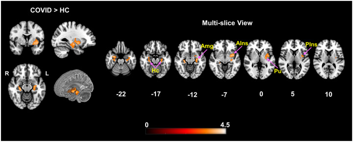
Results: The COVID group experienced significantly higher fatigue levels and GMV of this group was significantly higher within the Limbic System and Basal Ganglia when compared to healthy controls. Moreover, while a significant positive correlation was observed across the whole group between GMV and self-reported fatigue, COVID subjects showed stronger effects within the Posterior Cingulate, Precuneus and Superior Parietal Lobule .
Conclusion: Brain regions with GMV alterations in our analysis align with both single case acute patient reports and current group level neuroimaging findings. We also newly report a stronger positive correlation of GMV with fatigue among COVID survivors within brain regions associated with fatigue, indicating a link between structural abnormality and brain function in this cohort.
Figures

References
-
- (WHO) WHO. WHO Coronavirus (COVID-19) Dashboard 2022 [February/12/2022]. Available from: WHO Coronavirus (COVID-19) Dashboard.
-
- Kremer S, Lersy F, Anheim M, Merdji H, Schenck M, Oesterlé H, Bolognini F, Messie J, Khalil A, Gaudemer A, Carré S, Alleg M, Lecocq C, Schmitt E, Anxionnat R, Zhu F, Jager L, Nesser P, Mba YT, Hmeydia G, Benzakoun J, Oppenheim C, Ferré JC, Maamar A, Carsin-Nicol B, Comby PO, Ricolfi F, Thouant P, Boutet C, Fabre X, Forestier G, de Beaurepaire I, Bornet G, Desal H, Boulouis G, Berge J, Kazémi A, Pyatigorskaya N, Lecler A, Saleme S, Edjlali-Goujon M, Kerleroux B, Constans JM, Zorn PE, Mathieu M, Baloglu S, Ardellier FD, Willaume T, Brisset JC, Caillard S, Collange O, Mertes PM, Schneider F, Fafi-Kremer S, Ohana M, Meziani F, Meyer N, Helms J, Cotton F. Neurologic and neuroimaging findings in patients with COVID-19: A retrospective multicenter study. Neurology. 2020;95(13):e1868–e82. Epub 2020/07/19. doi: 10.1212/wnl.0000000000010112. - DOI - PubMed
-
- Keller E, Brandi G, Winklhofer S, Imbach LL, Kirschenbaum D, Frontzek K, Steiger P, Dietler S, Haeberlin M, Willms J, Porta F, Waeckerlin A, Huber M, Abela IA, Lutterotti A, Stippich C, Globas C, Varga Z, Jelcic I. Large and Small Cerebral Vessel Involvement in Severe COVID-19: Detailed Clinical Workup of a Case Series. Stroke. 2020;51(12):3719–22. Epub 2020/10/16. doi: 10.1161/strokeaha.120.031224. - DOI - PMC - PubMed
Publication types
Grants and funding
LinkOut - more resources
Full Text Sources