. 2022 Jan;63(1):61-74.
doi: 10.1111/epi.17130. Epub 2021 Nov 29.

Atlas of lesion locations and postsurgical seizure freedom in focal cortical dysplasia: A MELD study

Konrad Wagstyl  1 Kirstie Whitaker  2 Armin Raznahan  3 Jakob Seidlitz  4   5 Petra E Vértes  6 Stephen Foldes  7 Zachary Humphreys  7 Wenhan Hu  8 Jiajie Mo  8 Marcus Likeman  9 Shirin Davies  10   11 Matteo Lenge  12 Nathan T Cohen  13 Yingying Tang  14   15 Shan Wang  15   16 Mathilde Ripart  17 Aswin Chari  17   18 Martin Tisdall  17   18 Nuria Bargallo  19   20 Estefanía Conde-Blanco  19   20 Jose Carlos Pariente  20 Saül Pascual-Diaz  20 Ignacio Delgado-Martínez  21 Carmen Pérez-Enríquez  21 Ilaria Lagorio  22 Eugenio Abela  23 Nandini Mullatti  24 Jonathan O'Muircheartaigh  24 Katy Vecchiato  24 Yawu Liu  25 Maria Caligiuri  26 Ben Sinclair  27   28 Lucy Vivash  27   28 Anna Willard  27   28 Jothy Kandasamy  29 Ailsa McLellan  29 Drahoslav Sokol  29 Mira Semmelroch  30 Ane Kloster  31 Giske Opheim  31 Clarissa Yasuda  32 Kai Zhang  8 Khalid Hamandi  10   11 Carmen Barba  12 Renzo Guerrini  12 William Davis Gaillard  13 Xiaozhen You  13 Irene Wang  15 Sofía González-Ortiz  21 Mariasavina Severino  22 Pasquale Striano  22 Domenico Tortora  22 Reetta Kalviainen  25   33 Antonio Gambardella  26 Angelo Labate  26 Patricia Desmond  34 Elaine Lui  34 Terry O'Brien  27   28 Jay Shetty  29 Graeme Jackson  30 John S Duncan  35 Gavin P Winston  35   36 Lars Pinborg  31 Fernando Cendes  32 Judith Helen Cross  17   18 Torsten Baldeweg  17   18 Sophie Adler  17   18
Affiliations

Atlas of lesion locations and postsurgical seizure freedom in focal cortical dysplasia: A MELD study

Konrad Wagstyl et al. Epilepsia. 2022 Jan.

Erratum in

  • Erratum.
    [No authors listed] [No authors listed] Epilepsia. 2022 Apr;63(4):1018. doi: 10.1111/epi.17197. Epub 2022 Feb 28. Epilepsia. 2022. PMID: 35225361 Free PMC article. No abstract available.

Abstract

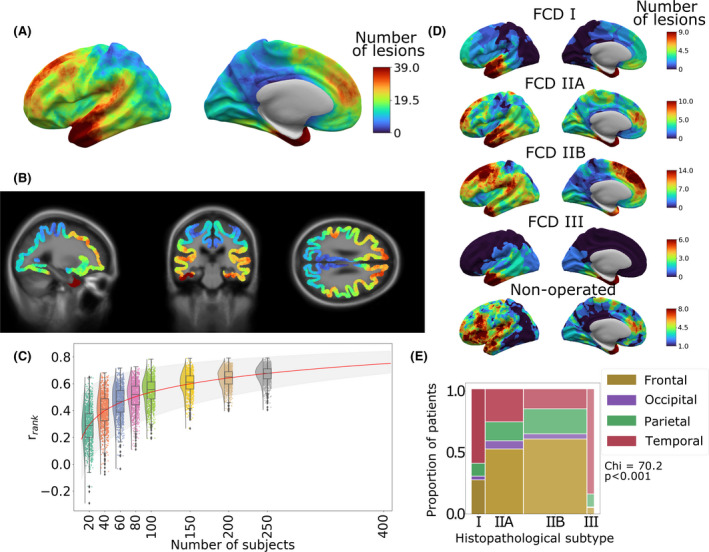
Objective: Drug-resistant focal epilepsy is often caused by focal cortical dysplasias (FCDs). The distribution of these lesions across the cerebral cortex and the impact of lesion location on clinical presentation and surgical outcome are largely unknown. We created a neuroimaging cohort of patients with individually mapped FCDs to determine factors associated with lesion location and predictors of postsurgical outcome.

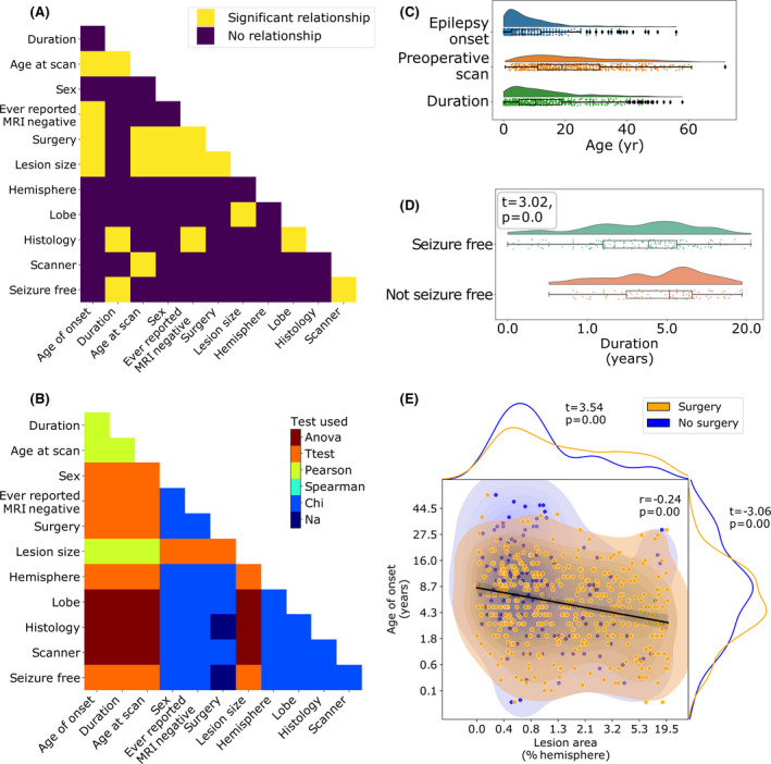
Methods: The MELD (Multi-centre Epilepsy Lesion Detection) project collated a retrospective cohort of 580 patients with epilepsy attributed to FCD from 20 epilepsy centers worldwide. Magnetic resonance imaging-based maps of individual FCDs with accompanying demographic, clinical, and surgical information were collected. We mapped the distribution of FCDs, examined for associations between clinical factors and lesion location, and developed a predictive model of postsurgical seizure freedom.

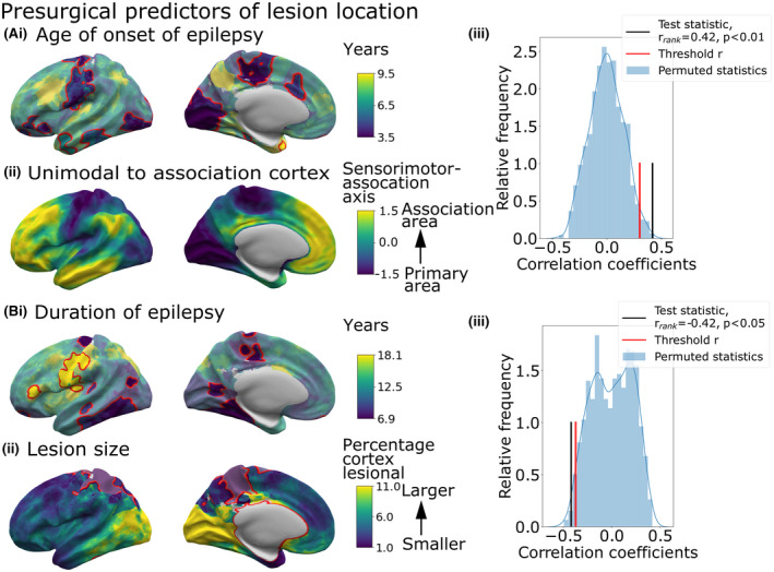
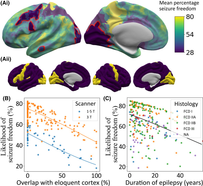
Results: FCDs were nonuniformly distributed, concentrating in the superior frontal sulcus, frontal pole, and temporal pole. Epilepsy onset was typically before the age of 10 years. Earlier epilepsy onset was associated with lesions in primary sensory areas, whereas later epilepsy onset was associated with lesions in association cortices. Lesions in temporal and occipital lobes tended to be larger than frontal lobe lesions. Seizure freedom rates varied with FCD location, from around 30% in visual, motor, and premotor areas to 75% in superior temporal and frontal gyri. The predictive model of postsurgical seizure freedom had a positive predictive value of 70% and negative predictive value of 61%.

Significance: FCD location is an important determinant of its size, the age at epilepsy onset, and the likelihood of seizure freedom postsurgery. Our atlas of lesion locations can be used to guide the radiological search for subtle lesions in individual patients. Our atlas of regional seizure freedom rates and associated predictive model can be used to estimate individual likelihoods of postsurgical seizure freedom. Data-driven atlases and predictive models are essential for evidence-based, precision medicine and risk counseling in epilepsy.

Keywords: MRI; drug-resistant epilepsy; focal cortical dysplasia; lesions; neurosurgery.


Conflict of interest statement

None of the authors has any conflict of interest to disclose.

Figures

FIGURE 1
FIGURE 1
Distribution of focal cortical dysplasia (FCD) lesions across the cerebral cortex. (A) All FCD lesion masks mapped to the left hemisphere of the template cortical surface. The distribution of FCDs across the cerebral cortex is nonuniform, with higher concentrations in the superior frontal sulcus, frontal pole, temporal pole, and superior temporal gyrus. (B) Three‐dimensional lesion likelihood atlas. Aggregated surface‐based lesion map values were normalized to between 0 and 1 and mapped back to the template magnetic resonance imaging volume. (C) Sample size required for consistent FCD lesion map. Rank correlation (y axis) was calculated by comparing the lesion map from a smaller cohort to a larger withheld cohort (n = 250). rrank increased with sample size. Predictive learning curve (red line) estimated that a stable map of lesion distribution requires a sample size of n = 400. (D) Distribution of FCD lesions according to histopathological subtype. (E) Distributions of lesions across cortical lobes within each FCD histopathological subtype. The width of bars indicates the relative numbers of patients. Temporal lobe lesions made up larger proportions of FCD Types I and III, whereas frontal lobe lesions were more likely to be FCD Types IIA and IIB
FIGURE 2
FIGURE 2
Presurgical factors associated with lesion location. Surface‐based maps show distribution of demographic variables according to lesion location. The color at each vertex represents the average variable value for patients with overlapping lesions. Vertices where the presence of a focal lesion was significantly associated with that variable are delineated in red. Factors significantly (p < .01) associated with lesion location were (Ai) age at epilepsy onset, (Bi) duration of epilepsy, and (Bii) lesion size. (Aiii) Correlation between the sensorimotor‐association axis of cortical organization (Aii) and the age at epilepsy onset (Ai) maps in comparison to spatially permuted maps. Lesions in primary areas were associated with a younger age at onset, whereas association areas had older ages of onset (rrank  = 0.39, pspin  < .01). (Biii) Correlation between the duration of epilepsy (Bi) and lesion size (Bii) maps in comparison to spatially permuted maps. Mean duration was significantly negatively correlated with the size of epilepsy lesion, where cortical areas with smaller lesions, for example, precentral and frontal areas, were associated with a longer duration of epilepsy, whereas areas with larger lesions, for example, occipital cortex, had shorter durations of epilepsy (rrank  = −0.42, pspin  < .05)
FIGURE 3
FIGURE 3
Effect of lesion location, duration of epilepsy, and histopathological subtype on seizure freedom. (Ai) Percentage of patients seizure‐free (%) according to lesion location across the cerebral cortex. Visual, motor, and premotor areas had a low percentage of seizure‐free patients (30%–40%). (Aii) Mask of eloquent cortex. (B) Impact of overlap of lesion with eloquent cortex and magnetic resonance imaging scanner field strength on likelihood of seizure freedom. (C) Impact of duration and histopathology on predicted percentage likelihood of seizure freedom. FCD, focal cortical dysplasia; NA, not available
FIGURE 4
FIGURE 4
Interrelationships between features. (A) Pairwise comparison of demographic and clinical features. Significant relationships after correction for multiple comparisons are shown in yellow. (B) Statistical test used for each pairwise comparison. (C) Distributions of age at epilepsy onset, age at magnetic resonance imaging (MRI) scan, and duration of epilepsy. (D) Duration of epilepsy is significantly associated with seizure freedom (t = −3.0, p < .001). Patients with longer durations of epilepsy are less likely to be seizure‐free. (E) Age at epilepsy onset, lesion size (as a percentage of the total hemisphere size), and seizure freedom are significantly associated. Larger lesions are associated with younger age at epilepsy onset (r = −0.24, p < .001) and are more likely to be operated on (t = 3.69, p < .001). Similarly, patients with a younger age at epilepsy onset are more likely to be operated on (t = −3.76, p < .001). Anova, analysis of variance; Na, not applicable

References

    1. US Institute of Medicine Committee on the Public Health Dimensions of the Epilepsies . Epilepsy across the spectrum: promoting health and understanding. England MJ, Liverman CT, Schultz AM, Strawbridge LM, editors. Washington, DC: National Academies Press; 2012. - PubMed
    1. Blümcke I, Thom M, Aronica E, Armstrong DD, Vinters HV, Palmini A, et al. The clinicopathologic spectrum of focal cortical dysplasias: a consensus classification proposed by an ad hoc Task Force of the ILAE Diagnostic Methods Commission. Epilepsia. 2011;52(1):158–74. - PMC - PubMed
    1. Blumcke I, Spreafico R, Haaker G, Coras R, Kobow K, Bien CG, et al. Histopathological findings in brain tissue obtained during epilepsy surgery. N Engl J Med. 2017;377(17):1648–56. - PubMed
    1. Lamberink HJ, Otte WM, Blümcke I, Braun KPJ, Aichholzer M, European Epilepsy Brain Bank writing group, study group , et al. Seizure outcome and use of antiepileptic drugs after epilepsy surgery according to histopathological diagnosis: a retrospective multicentre cohort study. Lancet Neurol. 2020;19(9):748–57. - PubMed
    1. Chassoux F, Devaux B, Landré E, Turak B, Nataf F, Varlet P, et al. Stereoelectroencephalography in focal cortical dysplasia: a 3D approach to delineating the dysplastic cortex. Brain. 2000;123(Pt 8):1733–51. - PubMed

Publication types

MeSH terms