. 2021 Dec 21;60(50):3829-3840.
doi: 10.1021/acs.biochem.1c00627. Epub 2021 Nov 30.

Second-Shell Amino Acid R266 Helps Determine N-Succinylamino Acid Racemase Reaction Specificity in Promiscuous N-Succinylamino Acid Racemase/ o-Succinylbenzoate Synthase Enzymes

Affiliations

Second-Shell Amino Acid R266 Helps Determine N-Succinylamino Acid Racemase Reaction Specificity in Promiscuous N-Succinylamino Acid Racemase/ o-Succinylbenzoate Synthase Enzymes

Dat P Truong et al. Biochemistry. .

Abstract

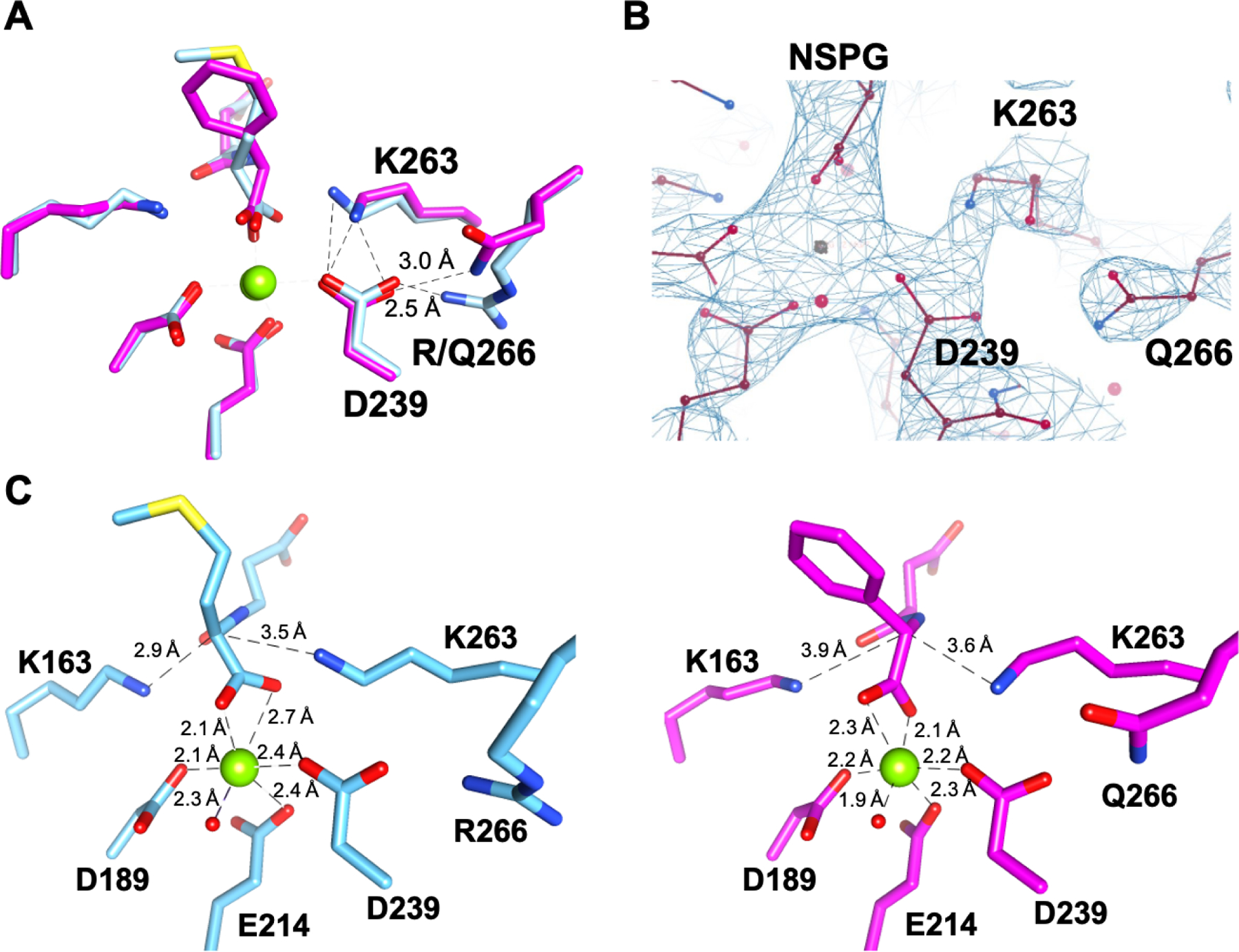
Catalytic promiscuity is the coincidental ability to catalyze nonbiological reactions in the same active site as the native biological reaction. Several lines of evidence show that catalytic promiscuity plays a role in the evolution of new enzyme functions. Thus, studying catalytic promiscuity can help identify structural features that predispose an enzyme to evolve new functions. This study identifies a potentially preadaptive residue in a promiscuous N-succinylamino acid racemase/o-succinylbenzoate synthase (NSAR/OSBS) enzyme from Amycolatopsis sp. T-1-60. This enzyme belongs to a branch of the OSBS family which includes many catalytically promiscuous NSAR/OSBS enzymes. R266 is conserved in all members of the NSAR/OSBS subfamily. However, the homologous position is usually hydrophobic in other OSBS subfamilies, whose enzymes lack NSAR activity. The second-shell amino acid R266 is close to the catalytic acid/base K263, but it does not contact the substrate, suggesting that R266 could affect the catalytic mechanism. Mutating R266 to glutamine in Amycolatopsis NSAR/OSBS profoundly reduces NSAR activity but moderately reduces OSBS activity. This is due to a 1000-fold decrease in the rate of proton exchange between the substrate and the general acid/base catalyst K263. This mutation is less deleterious for the OSBS reaction because K263 forms a cation-π interaction with the OSBS substrate and/or the intermediate, rather than acting as a general acid/base catalyst. Together, the data explain how R266 contributes to NSAR reaction specificity and was likely an essential preadaptation for the evolution of NSAR activity.


Conflict of interest statement

The authors declare no competing financial interest.
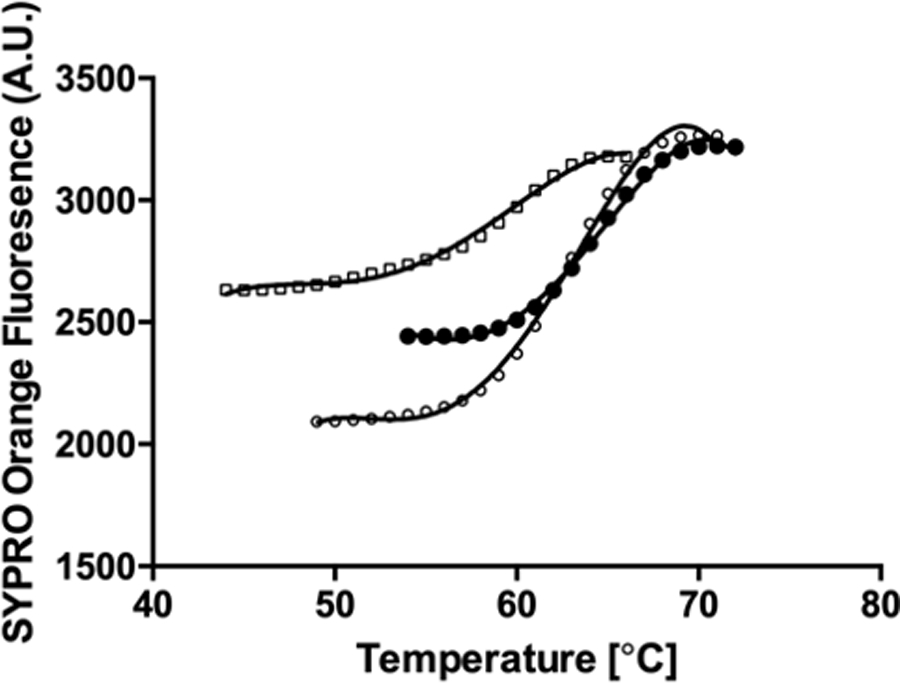
Figures

Figure 1.
Figure 1.
Phylogeny and sequence conservation in the OSBS family. A) The phylogeny of the OSBS family consists of several large subfamilies, which primarily correspond to the phylum from which the OSBS enzymes originated. NSAR activity is only found within the NSAR/OSBS subfamily (red). The width of the wedges in the phylogeny is proportional to the number of sequences included in that subfamily. Adapted with permission from ref. . Copyright 2012 American Chemical Society. B) Sequence logos showing differences in the conservation of amino acids at positions 261 and 266 among OSBS subfamilies. Sequence logos were generated by WebLogo. The letter size is proportional to the frequency of the amino acid at that position in the sequence alignment. Sequence numbering is relative to AmyNSAR/OSBS. N261 and R266 are highlighted in yellow and cyan, respectively. The catalytic K263 is highlighted in pink.
Figure 2.
Figure 2.
The mechanisms of the o-succinylbenzoate synthase (OSBS) and N-succinylamino acid racemase (NSAR) reactions. The divalent metal ion-stabilized enolate intermediate shared by the two reactions is shown in red. The atoms that are rearranged or lost during catalysis are shown in blue. The catalytic lysines shared by the two reactions are shown in magenta. N-succinylphenylglycine is shown as the substrate in the NSAR reaction, although characterized enzymes from the NSAR/OSBS subfamily can use a range of primarily hydrophobic N-succinylamino acids. Residues are numbered according to the sequence of AmyNSAR/OSBS.
Figure 3.
Figure 3.
Differential scanning fluorimetry of AmyNSAR/OSBS variants. The curves depict WT AmyNSAR/OSBS (closed circles), R266Q AmyNSAR/OSBS (open circles), and N261L AmyNSAR/OSBS (open squares).
Figure 4.
Figure 4.
pH-rate profiles of AmyNSAR/OSBS variants. (A) log(kcat/KM) vs. pH for the OSBS reaction of AmyNSAR/OSBS WT, N261L, and R266Q. (B) log(kcat/KM) vs. pH for the NSAR reaction of AmyNSAR/OSBS WT, N261L, and R266Q with D-NSPG. The same results were obtained with L-NSPG. (C) log(kcat) vs. pH of the NSAR reaction of AmyNSAR/OSBS WT, N261L, and R266Q. Data points above pH 9 are not shown for AmyNSAR/OSBS WT, because substrate saturation could not be achieved and thus kcat could not be determined.
Figure 5.
Figure 5.
R266Q specifically affects kex of K263. (A) Experimental scheme to measure the deuterium-hydrogen exchange rate, kex. The general base catalyst for each enantiomer is shown with curved arrows. The general acid catalyst could either be the same lysine, resulting in the same enantiomer (shown) or the lysine on the opposite side of the active site, resulting in the other enantiomer (not shown). (B) kex values measured for AmyNSAR/OSBS WT and NSPG. The structure of AmyNSAR/OSBS WT (PDB ID 1SJC; cyan) is shown, and L-NSPG (magenta) or D-NSPG (green) has been modeled into the structure. (C) kex values measured for AmyNSAR/OSBS R266Q (magenta) and NSPG. D-NSPG has been modeled into the experimental structure of AmyNSAR/OSBS R266Q.
Figure 6.
Figure 6.
Active site structure of AmyNSAR/OSBS R266Q. (A) Superimposed structures of wild-type and R266Q AmyNSAR/OSBS. Wild-type AmyNSAR/OSBS is bound to N-succinylmethionine and is shown in cyan (PDB ID 1SJC); AmyNSAR/OSBS R266Q is bound to N-succinylphenylglycine and is shown in magenta. The distance between R266 or Q266 and D239 are shown. Other dashed lines indicate distances of 3 Å or less among atoms of D239, K263, and R or Q266. Mg2+ is shown as a green sphere. (B) Electron density map showing the resolution of K263, Q266, D239 and other active site residues in AmyNSAR/OSBS R266Q. Mg2+ is shown as a gray sphere. (C) Comparison of metal ligands between wild-type AmyNSAR/OSBS bound to N-succinylmethionine (cyan; PDB ID 1SJC) and the R266Q mutant bound to N-succinylphenylglycine (magenta).

Similar articles

Cited by

References

    1. Brodkin HR, Novak WR, Milne AC, D’Aquino JA, Karabacak NM, Goldberg IG, Agar JN, Payne MS, Petsko GA, Ondrechen MJ, and Ringe D (2011) Evidence of the participation of remote residues in the catalytic activity of Co-type nitrile hydratase from Pseudomonas putida, Biochemistry 50, 4923–4935. - PMC - PubMed
    1. Blaisse MR, Fu B, and Chang MCY (2018) Structural and Biochemical Studies of Substrate Selectivity in Ascaris suum Thiolases, Biochemistry 57, 3155–3166. - PubMed
    1. Yang G, Hong N, Baier F, Jackson CJ, and Tokuriki N (2016) Conformational Tinkering Drives Evolution of a Promiscuous Activity through Indirect Mutational Effects, Biochemistry 55, 4583–4593. - PubMed
    1. Graf L, Craik CS, Patthy A, Roczniak S, Fletterick RJ, and Rutter WJ (1987) Selective alteration of substrate specificity by replacement of aspartic acid-189 with lysine in the binding pocket of trypsin, Biochemistry 26, 2616–2623. - PubMed
    1. Perona JJ, Hedstrom L, Rutter WJ, and Fletterick RJ (1995) Structural origins of substrate discrimination in trypsin and chymotrypsin, Biochemistry 34, 1489–1499. - PubMed

Publication types

MeSH terms