

Solitary jejunal diverticulum: a rare unexpected cause of chronic pneumoperitoneum
- PMID: 34846191
- PMCID: PMC10335030
- DOI: 10.1308/rcsann.2021.0233
Solitary jejunal diverticulum: a rare unexpected cause of chronic pneumoperitoneum
Abstract
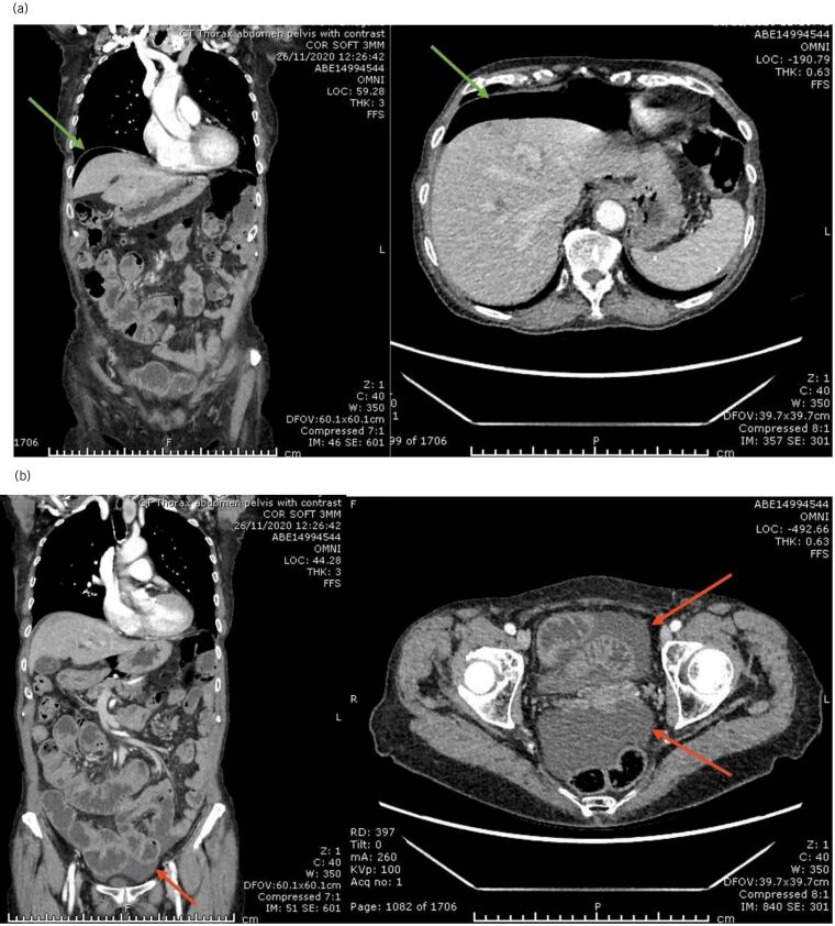
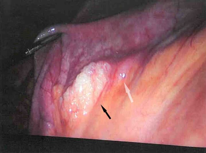
Jejunal diverticula are a rare acquired herniation of the mucosa and submucosa through the muscularis propria. They are asymptomatic in the majority of cases; however, they can present with non-specific abdominal symptoms and rarely complicate leading to acute abdomen. Perforation usually results in symptoms and signs of acute peritonitis and it is not an identifiable aetiology of chronic pneumoperitoneum. Computed tomography scanning may identify intestinal wall oedema, air bubbles travelling through the mesentery, free intra-abdominal air and/or fluid. Radiological diagnosis requires a high index of suspicion of such pathology. We report a case of an isolated jejunal diverticulum as a cause for aseptic chronic pneumoperitoneum.
Keywords: Intestinal perforations; Jejunal disease; Pneumoperitoneum; Unexpected.
Figures

Similar articles
-
A Case of Perforated Jejunal Diverticulum: An Unexpected Cause of Pneumoperitoneum in a Patient Presenting with an Acute Abdomen.Am J Case Rep. 2018 May 10;19:549-552. doi: 10.12659/AJCR.909131. Am J Case Rep. 2018. PMID: 29743472 Free PMC article.
-
Perforated isolated jejunal diverticula: a rare cause of acute abdomen.Clin J Gastroenterol. 2020 Oct;13(5):728-731. doi: 10.1007/s12328-020-01148-x. Epub 2020 Jun 10. Clin J Gastroenterol. 2020. Retraction in: Clin J Gastroenterol. 2021 Apr;14(2):697. doi: 10.1007/s12328-020-01266-6. PMID: 32524464 Retracted.
-
Perforation of jejunal diverticulum with ectopic pancreas.Clin J Gastroenterol. 2017 Apr;10(2):137-141. doi: 10.1007/s12328-017-0712-9. Epub 2017 Jan 19. Clin J Gastroenterol. 2017. PMID: 28102481
-
Perforation of jejunal diverticulum: case report and review of literature.Conn Med. 2000 Jan;64(1):7-10. Conn Med. 2000. PMID: 10697359 Review.
-
Small bowel diverticula in elderly patients: a case report and review article.BMC Surg. 2022 Mar 18;22(1):101. doi: 10.1186/s12893-022-01541-y. BMC Surg. 2022. PMID: 35303837 Free PMC article. Review.
References
-
- Syllaios A, Koutras A, Zotos PAet al. . Jejunal diverticulitis mimicking small bowel perforation: case report and review of the literature. Chirurgia (Romania). 2018; 113: 576–581. - PubMed
-
- Dunn V, Nelson JA. Jejunal diverticulosis and chronic pneumoperitoneum. Gastrointest Radiol 1979; 4: 165–168. - PubMed
Publication types
MeSH terms
LinkOut - more resources
Full Text Sources
Medical