. 2021 Dec 14;144(24):1940-1954.
doi: 10.1161/CIRCULATIONAHA.121.056414. Epub 2021 Nov 30.

Oxidized Phospholipids Promote NETosis and Arterial Thrombosis in LNK(SH2B3) Deficiency

Affiliations

Oxidized Phospholipids Promote NETosis and Arterial Thrombosis in LNK(SH2B3) Deficiency

Huijuan Dou et al. Circulation. .

Erratum in

Abstract

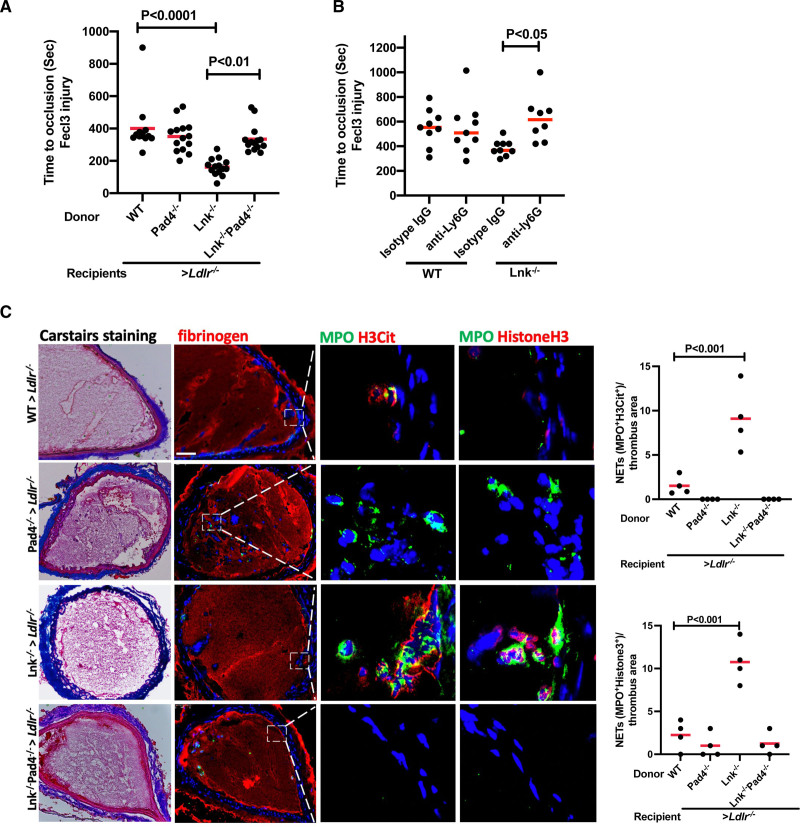
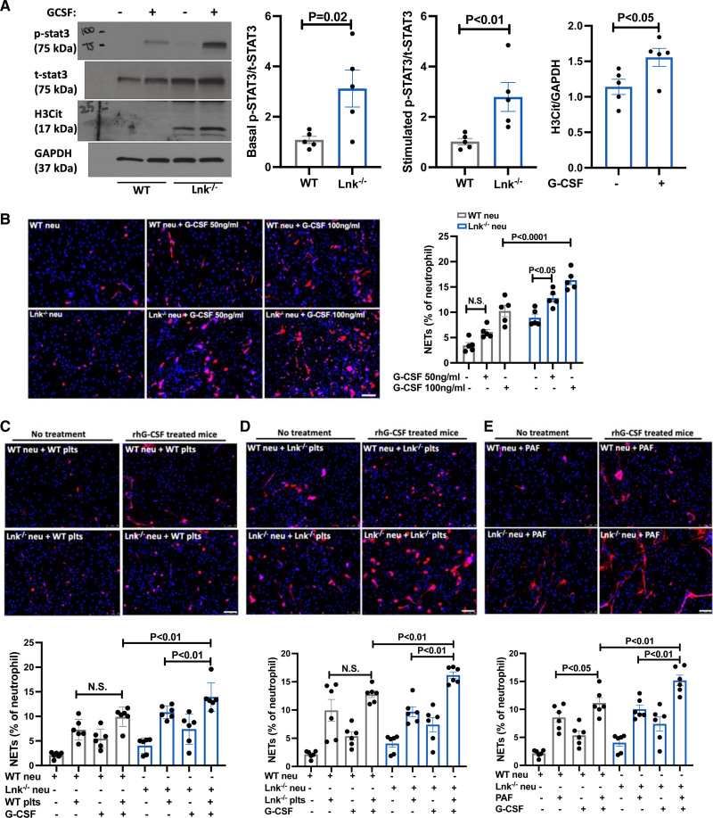
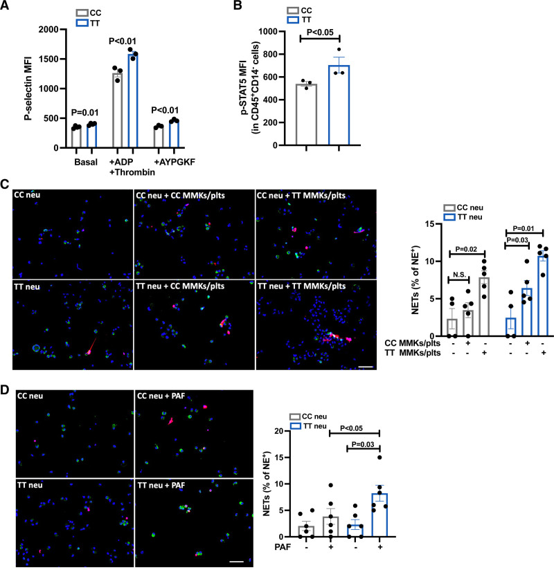
Background: LNK/SH2B3 inhibits Janus kinase/signal transducer and activator of transcription (JAK/STAT) signaling by hematopoietic cytokine receptors. Genome-wide association studies have shown association of a common single nucleotide polymorphism in LNK (R262W, T allele) with neutrophilia, thrombocytosis, and coronary artery disease. We have shown that LNK(TT) reduces LNK function and that LNK-deficient mice display prominent platelet-neutrophil aggregates, accelerated atherosclerosis, and thrombosis. Platelet-neutrophil interactions can promote neutrophil extracellular trap (NET) formation. The goals of this study were to assess the role of NETs in atherosclerosis and thrombosis in mice with hematopoietic Lnk deficiency.

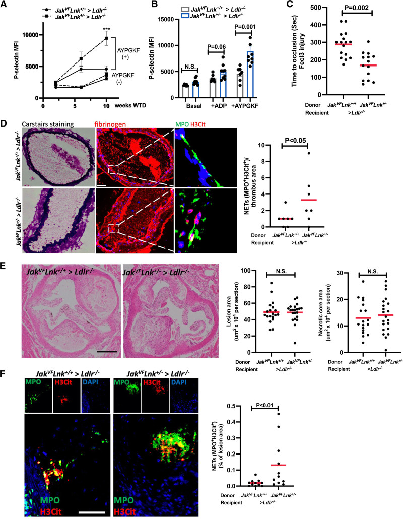
Methods: We bred mice with combined deficiency of Lnk and the NETosis-essential enzyme PAD4 (peptidyl arginine deiminase 4) and transplanted their bone marrow into Ldlr-/- mice. We evaluated the role of LNK in atherothrombosis in humans and mice bearing a gain of function variant in JAK2 (JAK2V617F).

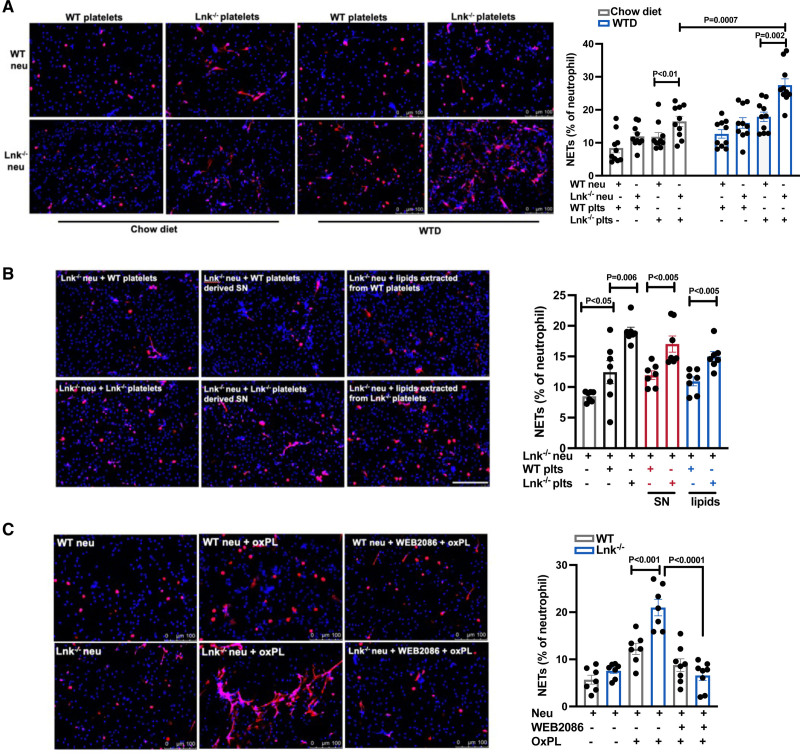
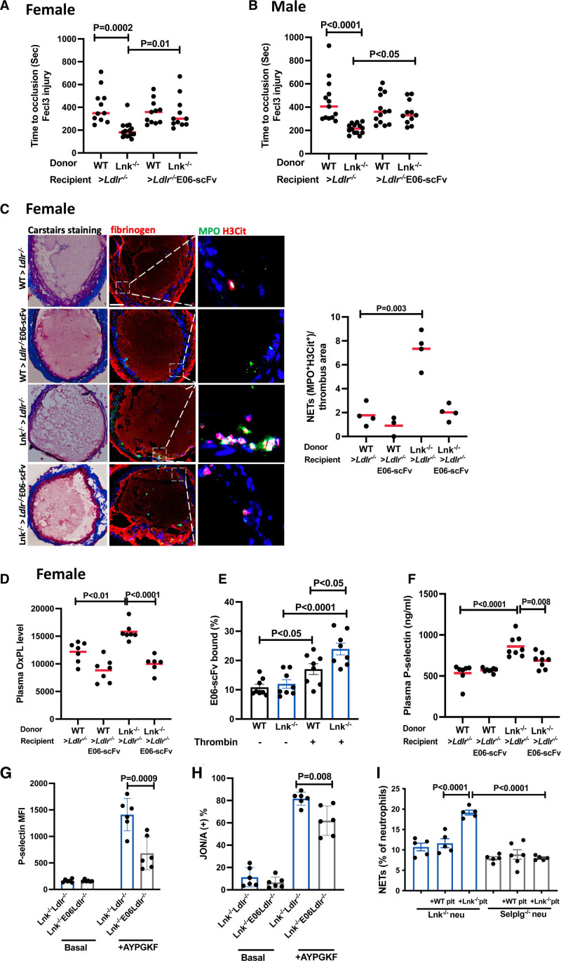
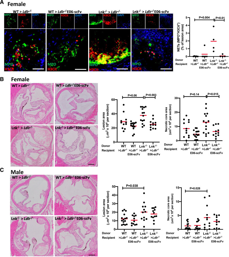
Results: Lnk-deficient mice displayed accelerated carotid artery thrombosis with prominent NETosis that was completely reversed by PAD4 deficiency. Thrombin-activated Lnk-/- platelets promoted increased NETosis when incubated with Lnk-/- neutrophils compared with wild-type platelets or wild-type neutrophils. This involved increased surface exposure and release of oxidized phospholipids (OxPL) from Lnk-/- platelets, as well as increased priming and response of Lnk-/- neutrophils to OxPL. To counteract the effects of OxPL, we introduced a transgene expressing the single-chain variable fragment of E06 (E06-scFv). E06-scFv reversed accelerated NETosis, atherosclerosis, and thrombosis in Lnk-/- mice. We also showed increased NETosis when human induced pluripotent stem cell-derived LNK(TT) neutrophils were incubated with LNK(TT) platelet/megakaryocytes, but not in isogenic LNK(CC) controls, confirming human relevance. Using data from the UK Biobank, we found that individuals with the JAK2VF mutation only showed increased risk of coronary artery disease when also carrying the LNK R262W allele. Mice with hematopoietic Lnk+/- and Jak2VF clonal hematopoiesis showed accelerated arterial thrombosis but not atherosclerosis compared with Jak2VFLnk+/+ controls.

Conclusions: Hematopoietic Lnk deficiency promotes NETosis and arterial thrombosis in an OxPL-dependent fashion. LNK(R262W) reduces LNK function in human platelets and neutrophils, promoting NETosis, and increases coronary artery disease risk in humans carrying Jak2VF mutations. Therapies targeting OxPL may be beneficial for coronary artery disease in genetically defined human populations.

Keywords: clonal hematopoiesis; coronary artery disease; extracellular traps; phospholipids; thrombosis.


Figures

Figure 1.
Figure 1.
Increased neutrophil extracellular traps and arterial thrombosis in Lnk–/– mice are reversed by PAD4 deficiency. A, FeCl3-induced carotid artery occlusion in wild-type (WT), Lnk–/–, Pad4–/–, and Lnk–/– Pad4–/– bone marrow female Ldlr–/– recipient mice fed Western-type diet for 10 weeks; 2-way analysis of variance. B, Time to occlusion measured in WT and Lnk–/– bone marrow recipient mice after 48 hours of treatment with 100 µg anti-Ly6G or isotype control antibody; 2-way analysis of variance. C, Representative images of Carstairs, fibrinogen (red), or neutrophil extracellular trap (NET) staining in carotid artery thrombi of WT, Lnk–/–, Pad4–/–, and Lnk–/–Pad4–/– bone marrow female Ldlr–/– recipients fed Western-type diet for 10 weeks. NETs were validated using MPO (green) with citrullinated histone (H3Cit; red) or MPO (green) with histone 3 (red). NETs shown were from the same area as in fibrinogen staining on consecutive sections. Quantification of NETs (n=4 per group); 2-way analysis of variance. Each point represents 1 mouse. Scale bar, 50 µm. Data are expressed as mean (red bar) ±SEM. IgG indicates immunoglobulin G.
Figure 2.
Figure 2.
Platelet-released platelet-activating factor–like oxidized phospholipid induces neutrophil extracellular traps in Lnk–/– neutrophils through platelet-activating factor receptor. A, Platelets activated with 1 nmol/L thrombin were incubated with neutrophils at a ratio=20:1 for 4 hours. Representative images of NETosis. Red, Citrullinated histone (H3Cit); blue, DAPI. Neutrophil extracellular trap (NET) numbers were normalized to total neutrophil number; 2-way analysis of variance. Scale bar,100 µm. B, Lnk–/– neutrophils were stimulated with wild-type (WT) or Lnk–/– platelets, platelet-derived supernatant, or platelet supernatant–derived lipids for 4 hours. Red, H3Cit; blue, DAPI; n=7. C, Neutrophils were preincubated with WEB2086 (4 µmol/L) for 30 minutes followed by 3 hours oxidized phospholipid (OxPL; 20 µg/mL) treatment and then stained with H3Cit and DAPI (n=7). Quantification of NETs; 2-way analysis of variance. Scale bar, 100 µm. Data are expressed as mean±SEM. SN indicates supernatant; and WTD, Western-type diet.
Figure 3.
Figure 3.
E06-scFv reverses increased neutrophil extracellular traps and accelerated thrombosis in Lnk–/– mice. FeCl3-induced carotid artery occlusion in Western-type diet–fed (10 weeks) female (A) or male (B) recipients of designated genotype as indicated; 2-way analysis of variance. C, Representative images of Carstairs, fibrinogen, or neutrophil extracellular trap (NET) staining in carotid artery thrombi of the female recipients as in A. NETs were validated using MPO (green) with citrullinated histone (H3Cit; red). NETs shown were from the same area as in fibrinogen staining on consecutive sections. Quantification of NETs in carotid artery thrombi of the female recipients (n=4 per group); 2-way analysis of variance. Scale bar, 50 µm. Each point represents 1 mouse. D, Plasma oxidized phospholipid (OxPL) level was assessed in the female recipients as in A (n=6–7). E, E06-scFv binding to the basal or activated platelets (n=8). F, Plasma P-selectin level was assessed by enzyme-linked immunosorbent assay in the female recipients as in A (n=8). Surface P-selectin (G) and active integrin αIIbβ3 (JON/A; H) levels on platelets with and without AYPGKF (50 µmol/L) stimulation (n=6); 2-way analysis of variance. I, Quantification of NETs from wild-type (WT) or Selplg–/– neutrophils stimulated by activated WT or Lnk–/– platelets (n=5); 2-way analysis of variance. Data are expressed as mean (red bar) ±SEM.
Figure 4.
Figure 4.
E06-scFv reverses increased neutrophil extracellular traps and atherosclerosis in Lnk–/– mice. A, Representative images showing staining for MPO (green) and citrullinated histone (H3Cit; red) with DAPI (blue) in aortic root lesions of wild-type (WT) or Lnk–/– bone marrow female Ldlr–/– or Ldlr–/– E06-scFv recipients fed with Western-type diet for 10 weeks. The graph quantitates neutrophil extracellular traps (NETs; MPO+H3Cit+) in lesions; 2-way analysis of variance. Scale bar, 50 µm. B and C, Representative hematoxylin & eosin–stained aortic root lesions and quantification of lesion area and necrotic core area in female (B) and male (C) Ldlr–/– and Ldlr–/– E06-scFv recipients after 10 weeks of Western-type diet (n=15–20); 2-way analysis of variance. Scale bar, 500 µm. Data are expressed as mean (red bar) ±SEM.
Figure 5.
Figure 5.
Granulocyte colony-stimulating factor promotes NETosis in LNK-deficient mice. A, Wild-type (WT) or Lnk–/– mice were intraperitoneally injected with vehicle or 2.5 μg recombinant human (rh) granulocyte colony-stimulating factor (G-CSF) for 15 minutes. p-STAT3 and citrullinated histone (H3Cit) levels in neutrophils isolated from WT or Lnk–/– mice were determined and quantified by Western analysis (n=5). Unpaired t test. B, WT or Lnk–/– neutrophils were stimulated with G-CSF for 3 hours. The cells were then stained with H3Cit (red) and DAPI and neutrophil extracellular traps (NETs) were quantified (n=5). C through E, WT or Lnk–/– mice were treated with IP injection of vehicle or rhG-CSF (2.5 μg) for 3 days; neutrophils from vehicle or rhG-CSF–treated WT or Lnk–/– mice were isolated and stimulated with WT platelets (C), Lnk–/– platelets (D), or platelet-activating factor (E) for 4 hours. NETs were quantified (n=6); 2-way analysis of variance. Scale bar, 50 µm. Data are expressed as mean±SEM.
Figure 6.
Figure 6.
Increased platelet activation, STAT signaling, and NETosis in human induced pluripotent stem cell–derived LNK(TT) cells. A, Surface P-selectin level on megakaryocytes (MMKs)/platelets with and without ADP (20 µmol/L) and thrombin (1 U/mL) or AYPGKF (50 µmol/L) stimulation from day 15 LNK(TT) and LNK(CC) cells (n=3). Unpaired t test. B, p-STAT5 level was analyzed in day 13 CD45+CD14 LNK(TT) and LNK(CC) cells (n=3). Unpaired t test. C, MMKs/platelets activated with 1 nmol/L thrombin were incubated with LNK(TT) and LNK(CC) neutrophils for 4 hours. Representative images of NETosis. Green, Neutrophil elastase; red, citrullinated histone (H3Cit); blue, DAPI. Neutrophil extracellular trap (NET) numbers were normalized to neutrophil elastase positive cell numbers; 2-way analysis of variance. Scale bar, 50 µm. D, LNK(TT) and LNK(CC) neutrophils were stimulated with 5 µmol/L platelet-activating factor (PAF) for 3 hours and quantified for NETs (n=6); 2-way analysis of variance. Scale bar, 50 µm. Data are expressed as mean±SEM.
Figure 7.
Figure 7.
Increased neutrophil extracellular traps and accelerated thrombosis in Jak2VFLnk+/– mice. A, Surface P-selectin level on platelets in whole blood with and without AYPGKF (50 µmol/L) stimulation from mice after 2, 6, and 10 weeks Western-type diet (WTD; n=6–7); 2-way analysis of variance. *P<0.05, **P<0.01, ***P<0.001. B, Surface P-selectin level on platelets in whole blood with and without ADP (20 µmol/L) and AYPGKF (50 µmol/L) after 10 weeks WTD feeding (n=7–8). C, FeCl3 induced carotid occlusion in bone marrow Ldlr–/– recipient mice (n=15–17). D, Representative images of Carstairs, fibrinogen, or neutrophil extracellular trap (NET) staining in thrombi of the recipients. NETs were validated using MPO (green) with citrullinated histone (H3Cit; red). Quantification of NETs in thrombi of the recipients (n=4 per group). Unpaired t test. Scale bar, 50 µm. Each point represents 1 mouse. E, Representative H&E-stained aortic root lesions and quantification of lesion area and necrotic core area in Ldlr–/– recipients after 10 weeks of WTD (n=15–17). Scale bar, 500 µm. F, Representative images showing staining for MPO (green) and citrullinated histone (H3Cit; red) with DAPI (blue) in aortic root lesions of Ldlr–/– recipients fed with WTD for 10 weeks and quantification of NETs (MPO+H3Cit+) in lesions. Unpaired t test. Scale bar, 50 µm.

References

    1. Deloukas P, Kanoni S, Willenborg C, Farrall M, Assimes TL, Thompson JR, Ingelsson E, Saleheen D, Erdmann J, Goldstein BA, et al. ; CARDIoGRAMplusC4D Consortium; DIAGRAM Consortium; CARDIOGENICS Consortium; MuTHER Consortium; Wellcome Trust Case Control Consortium. Large-scale association analysis identifies new risk loci for coronary artery disease. Nat Genet. 2013; 45:25–33. doi: 10.1038/ng.2480 - PMC - PubMed
    1. Gudbjartsson DF, Bjornsdottir US, Halapi E, Helgadottir A, Sulem P, Jonsdottir GM, Thorleifsson G, Helgadottir H, Steinthorsdottir V, Stefansson H, et al. Sequence variants affecting eosinophil numbers associate with asthma and myocardial infarction. Nat Genet. 2009; 41:342–347. doi: 10.1038/ng.323 - PubMed
    1. Barrett JC, Clayton DG, Concannon P, Akolkar B, Cooper JD, Erlich HA, Julier C, Morahan G, Nerup J, Nierras C, et al. ; Type 1 Diabetes Genetics Consortium. Genome-wide association study and meta-analysis find that over 40 loci affect risk of type 1 diabetes. Nat Genet. 2009; 41:703–707. doi: 10.1038/ng.381 - PMC - PubMed
    1. Dubois PC, Trynka G, Franke L, Hunt KA, Romanos J, Curtotti A, Zhernakova A, Heap GA, Adány R, Aromaa A, et al. Multiple common variants for celiac disease influencing immune gene expression. Nat Genet. 2010; 42:295–302. doi: 10.1038/ng.543 - PMC - PubMed
    1. Bersenev A, Wu C, Balcerek J, Tong W. Lnk controls mouse hematopoietic stem cell self-renewal and quiescence through direct interactions with JAK2. J Clin Invest. 2008; 118:2832–2844. doi: 10.1172/JCI35808 - PMC - PubMed

Publication types