

doi: 10.3324/haematol.2021.278779.
Predicting risk of progression in relapsed multiple myeloma using traditional risk models, focal lesion assessment with PET-CT and minimal residual disease status
Affiliations
- PMID: 34847659
- PMCID: PMC8634177
- DOI: 10.3324/haematol.2021.278779
Item in Clipboard
Predicting risk of progression in relapsed multiple myeloma using traditional risk models, focal lesion assessment with PET-CT and minimal residual disease status
Haematologica.
.
No abstract available
Figures
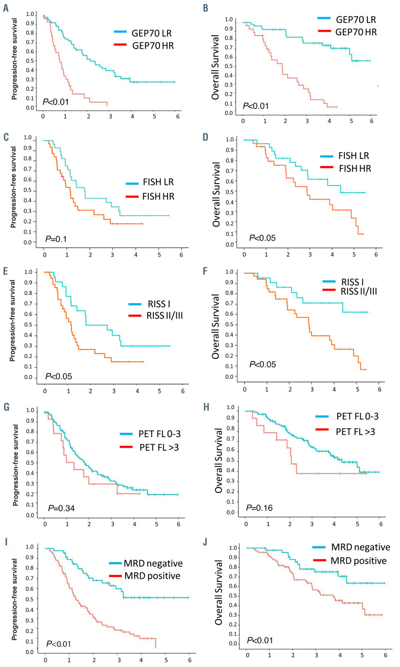
Reassessment of traditional risk factors in first multiple myeloma relapse shows improved prognostic accuracy compared to their evaluation at diagnosis (shown in the Online Supplementary Appendix). GEP70 high-risk (HR) patients at relapse had significant worse progression-free survival (PFS) (A), and OS (B) compared to low-risk (LR) patients, P<0.01. HR fluorescence in situ hybridization (FISH) included translocation t14;16, t4;14 and del 17p and showed worse PFS (P=0.1) (C) and OS (P<0.05) (D), compared to patients with LR FISH. Assessment of revised International Staging System (RISS) at relapse showed significant worse PFS, E, and OS, F, for patients with RISS 2+3 compared to patients with RISS stage I. The presence of >3 focal lesions by positron emission tomography and computed tomography (PET CT) at relapse was associated with worse PFS (H) and OS (I) in first relapse. The results were not significant, likely due to the relative small patient number. Achievement of minimal residual disease (MRD) negativity after first relapse was a powerful marker for significantly improved PFS (J) and OS (K).
References
-
- Kristinsson SY, Anderson WF, Landgren O. Improved long-term survival in multiple myeloma up to the age of 80 years. Leukemia. 2014; 28(6):1346-1348. - PubMed
-
- Schinke M, Ihorst G, Duyster J, Wasch R, Schumacher M, Engelhardt M. Risk of disease recurrence and survival in patients with multiple myeloma: a German Study Group analysis using a conditional survival approach with long-term follow-up of 815 patients. Cancer. 2020; 126(15):3504-3515. - PubMed
-
- Hillengass J, Usmani S, Rajkumar SV, et al. International myeloma working group consensus recommendations on imaging in monoclonal plasma cell disorders. Lancet Oncol. 2019;20(6):e302-e312. - PubMed
Publication types
MeSH terms
Substances
Grants and funding
LinkOut - more resources
Full Text Sources
Medical
