Review
. 2022 Jan:162:105574.
doi: 10.1016/j.nbd.2021.105574. Epub 2021 Nov 27.

Synaptic pathology in Huntington's disease: Beyond the corticostriatal pathway

Affiliations
Review

Synaptic pathology in Huntington's disease: Beyond the corticostriatal pathway

Joshua Barry et al. Neurobiol Dis. 2022 Jan.

Abstract

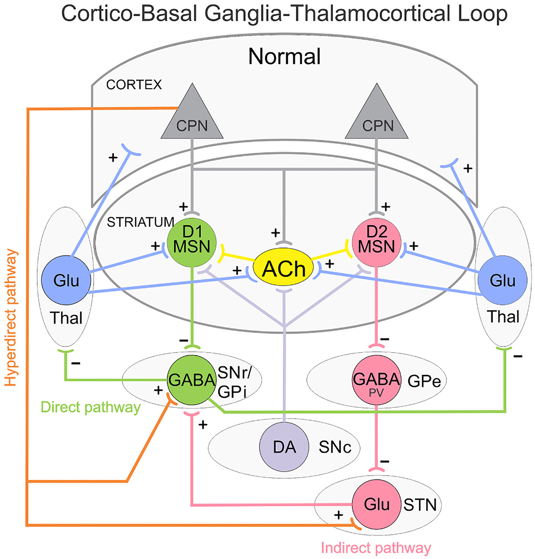
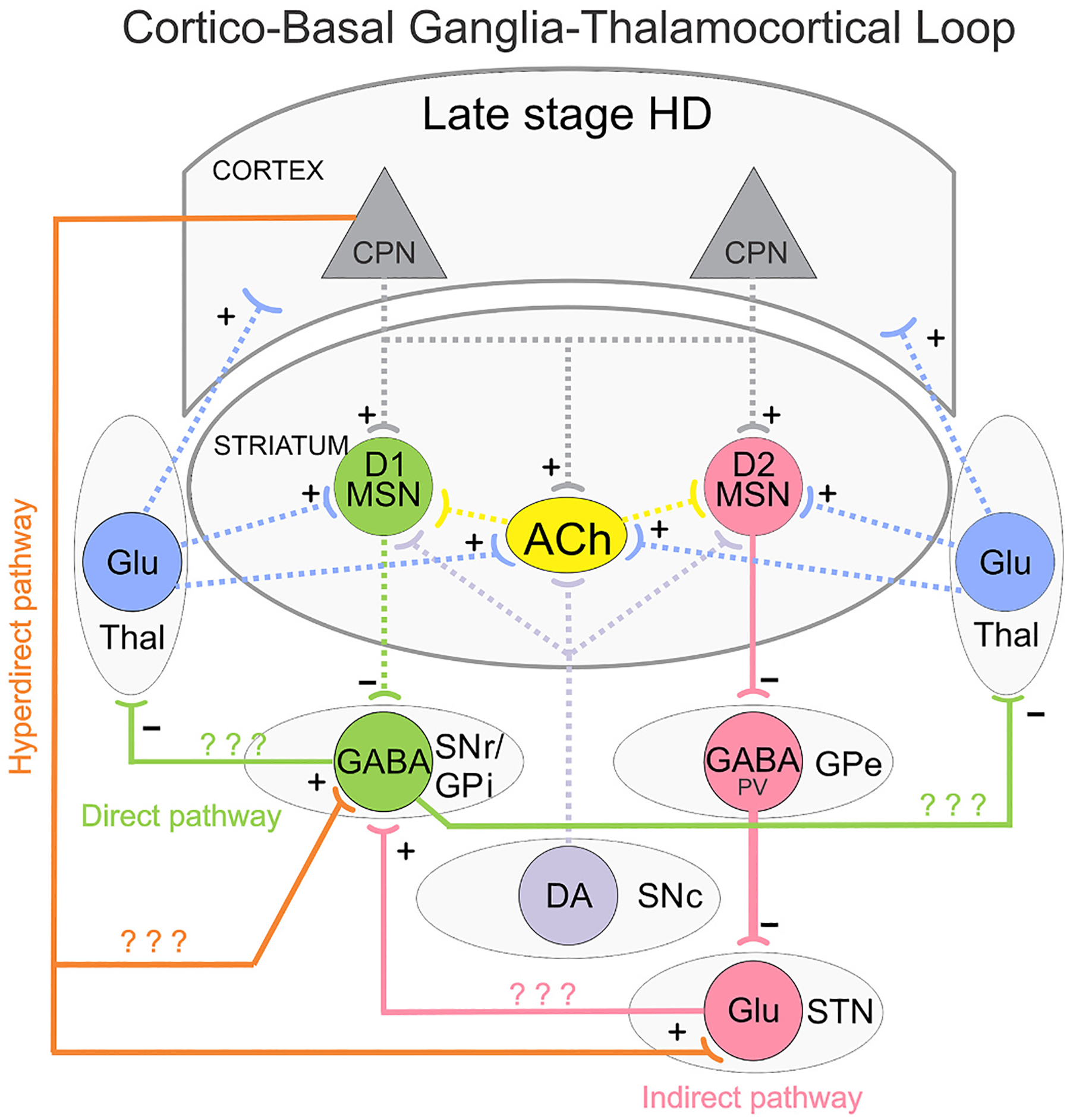
Huntington's disease (HD) is a heritable, fatal neurodegenerative disorder caused by a mutation in the Huntingtin gene. It is characterized by chorea, as well as cognitive and psychiatric symptoms. Histopathologically, there is a massive loss of striatal projection neurons and less but significant loss in other areas throughout the cortico-basal ganglia-thalamocortical (CBGTC) loop. The mutant huntingtin protein has been implicated in numerous functions, including an important role in synaptic transmission. Most studies on anatomical and physiological alterations in HD have focused on striatum and cerebral cortex. However, based on recent CBGTC projectome evidence, the need to study other pathways has become increasingly clear. In this review, we examine the current status of our knowledge of morphological and electrophysiological alterations of those pathways in animal models of HD. Based on recent studies, there is accumulating evidence that synaptic disconnection, particularly along excitatory pathways, is pervasive and almost universal in HD, thus supporting a critical role of the huntingtin protein in synaptic transmission.

Keywords: Basal ganglia; Disconnection; Genetic models; Huntington's disease; Synaptic activity.


Figures

Fig. 1.
Fig. 1.
Simplified diagram of the cortico-basal ganglia-thalamocortical (CBGTC) loop. Striatal medium-sized spiny neurons (MSNs) expressing dopamine (DA) D1 receptors receive glutamatergic (Glu) inputs (+) from cortical pyramidal neurons (CPNs) and project to the substantia nigra pars reticulata (SNr) as well as the internal segment of the globus pallidus (GPi). This is called the direct pathway and promotes movement. MSNs expressing DA D2 receptors also receive inputs from CPNs and project to the external segment of the globus pallidus (GPe). The GPe, in turn, projects to the subthalamic nucleus (STN) and this to the SNr/GPi. This is known as the indirect pathway and counteracts movement. GABA (−) outputs from SNr/GPi project to the thalamus (Thal), which in turn, sends Glu inputs (+) back to the cortex thus completing the CBGTC loop. Both D1 and D2 MSNs also receive afferents from the substantia nigra pars compacta (SNc) and Thal. A hyperdirect pathway connects the CPNs of the cortex directly to the STN. In addition, a newly described pathway from CPNs to SNr GABAergic neurons is illustrated. Several classes of interneurons, including acetylcholine (ACh) interneurons, exert control over the MSNs. For the sake of simplicity, GABAergic interneurons and some additional synaptic pathways are not illustrated.
Fig. 2.
Fig. 2.
Simplified diagram of the CBGTC loop in late stage HD. Glu inputs (+) from CPNs are reduced onto both direct (D1) and indirect (D2) pathway MSNs. GABA (−) outputs from D1 MSNs are reduced onto SNr/GPi neurons, dis-inhibiting these neurons and promoting hypokinesia. GABA (−) outputs from D2 MSNs are unchanged or decreased, but reduced GABA reuptake disrupts firing patterns of PV-expressing GABAergic neurons in the GPe and facilitate bursting activity. This, in conjunction with reduced cortical input through the hyperdirect pathway, may reduce STN firing. Question marks indicate that alterations of these pathways in HD remain unknown or studies are inconclusive.

References

    1. Akopian G, Barry J, Cepeda C, Levine MS, 2016. Altered membrane properties and firing patterns of external globus pallidus neurons in the R6/2 mouse model of Huntington’s disease. J. Neurosci. Res 94, 1400–1410. - PMC - PubMed
    1. Albin RL, Reiner A, Anderson KD, Dure LS, Handelin B, et al., 1992. Preferential loss of striato-external pallidal projection neurons in presymptomatic Huntington’s disease. Ann. Neurol 31, 425–430. - PubMed
    1. Alexander GE, DeLong MR, Strick PL, 1986. Parallel organization of functionally segregated circuits linking basal ganglia and cortex. Annu. Rev. Neurosci 9, 357–381. - PubMed
    1. Andre VM, Cepeda C, Fisher YE, Huynh M, Bardakjian N, et al., 2011. Differential electrophysiological changes in striatal output neurons in Huntington’s disease. J. Neurosci 31, 1170–1182. - PMC - PubMed
    1. Aoki S, Smith JB, Li H, Yan X, Igarashi M, et al., 2019. An open cortico-basal ganglia loop allows limbic control over motor output via the nigrothalamic pathway. eLife 8. - PMC - PubMed

Publication types

MeSH terms

Substances