. 2021 Dec;41(12):5919-5933.
doi: 10.21873/anticanres.15411.

Effect of Diallyl Trisulfide on TNF-α-induced CCL2/MCP-1 Release in Genetically Different Triple-negative Breast Cancer Cells

Affiliations

Effect of Diallyl Trisulfide on TNF-α-induced CCL2/MCP-1 Release in Genetically Different Triple-negative Breast Cancer Cells

Konan J W Kanga et al. Anticancer Res. 2021 Dec.

Abstract

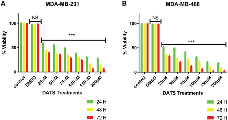
Background/aim: Diallyl trisulfide (DATS) has been shown to prevent and inhibit breast carcinogenesis. CCL2/MCP-1 has been shown to play a significant role in breast cancer. This study explored DATS efficacy on triple-negative breast cancer (TNBC) cells.

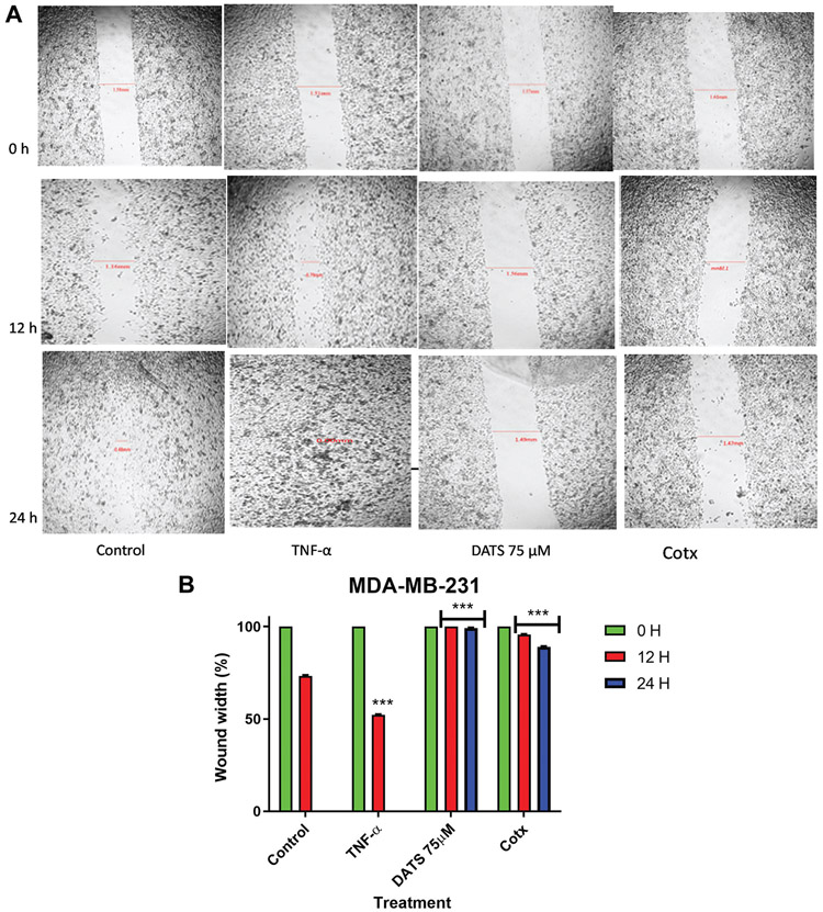
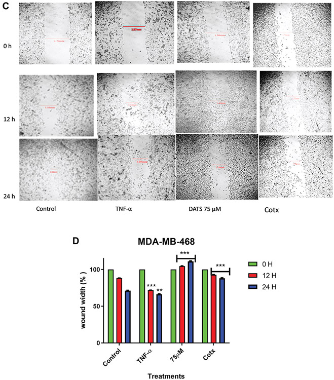
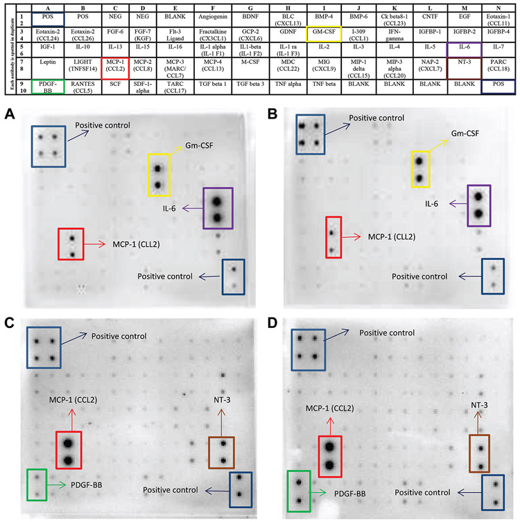
Materials and methods: DATS efficacy on TNF-α induced TNBC cells were examined via trypan blue exclusion test, wound-healing assay, human cytokine arrays, ELISA, and RT-PCR.

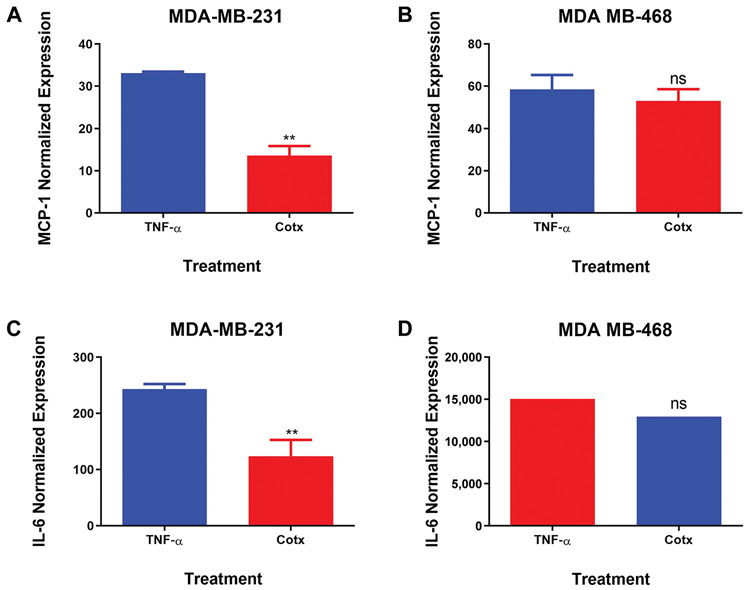
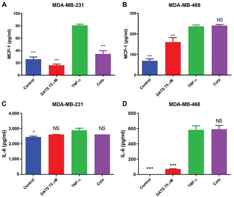
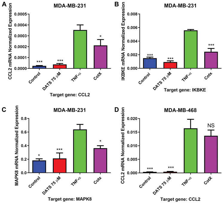
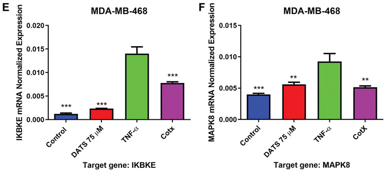
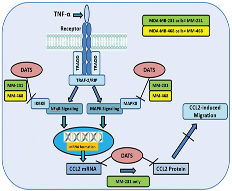
Results: DATS significantly induced cell death and inhibited cell migration. Expression of CCL2/MCP-1, IL-6, PDGF-BB, NT-3, and GM-CSF in TNF-α-treated cells increased. However, DATS significantly decreased the expression of CCL2/MCP-1 in TNF-α-treated MDA-MB-231 but not in MDA-MB-468 cells. DATS significantly down-regulated mRNA expression of IKBKE and MAPK8 in both cell lines, indicating a possible effect in genes involved in the NF-κB and MAPK signaling.

Conclusion: DATS may have a role in TNBC therapy and prevention by targeting CCL2.

Keywords: Breast cancer; CCL2; IKBKE; MAPK8; MCP-1; diallyl trisulfide; tumor necrosis factor alpha.


Conflict of interest statement

Conflicts of Interest

The Authors declare no competing interests concerning this study.

Figures

Figure 1.
Figure 1.
The effect of DATS on viability oϕ MDA-MB-231 (A) and MDA-MB-468 (B) TNBC cells. MDA-MB-231 and MDA-MB-468 were treated with various concentrations (0-200 μM) of DATS for 24, 48, and 72 h. The effect of DATS was both dose- and -time-dependent. Treatment with a low dose of 25 μM of DATS significantly decreased cell viability compared to control. DMSO treatment showed no cytotoxicity at any time point. The lethal concentration 50 (LC50) value was acquired from GraphPad Prism using mean values of data points at the previously mentioned concentrations. All experiments were performed at least three times with n=3. The data are presented as the mean±SEM. Statistically significant differences between control vs. treatments were evaluated by a one-way ANOVA, followed by’ ‘Dunnett’s multiple comparison test or Student’s t-test to compare the results between the two cell lines with ***p<0.001.
Figure 2.
Figure 2.
The effect of DATS on TNF-α induced MDA-MB-231 and MDA-MB-468 breast cancer cells. A wound-healing assay was used to examine the effect of diallyl trisulfide (DATS) on cell migration in TNF-α induced MDA-MB-231 and MDA-MB-468 breast cancer cells (A and C) at various time points (0, 12, and 24 h). Wound healing assay representative image and quantification of MDA-MB-231 and MDA-MB-468 cell migration with a magnification of 40X. (B and D) Wound width % was statistically analyzed. At least 3 independent experiments were performed with n=3. The data are presented as the mean±S.E.M. Statistically significant differences between control vs. treatments were evaluated by a one-way ANOVA, followed by Dunnett’s multiple comparison test with **p<0.01 and ***p<0.001.
Figure 2.
Figure 2.
The effect of DATS on TNF-α induced MDA-MB-231 and MDA-MB-468 breast cancer cells. A wound-healing assay was used to examine the effect of diallyl trisulfide (DATS) on cell migration in TNF-α induced MDA-MB-231 and MDA-MB-468 breast cancer cells (A and C) at various time points (0, 12, and 24 h). Wound healing assay representative image and quantification of MDA-MB-231 and MDA-MB-468 cell migration with a magnification of 40X. (B and D) Wound width % was statistically analyzed. At least 3 independent experiments were performed with n=3. The data are presented as the mean±S.E.M. Statistically significant differences between control vs. treatments were evaluated by a one-way ANOVA, followed by Dunnett’s multiple comparison test with **p<0.01 and ***p<0.001.
Figure 3.
Figure 3.
Comparative effect of diallyl trisulfide (DATS) on cytokine expression in TNF-alpha induced MDA-MB-231 and MDA-MB-468 breast cancer cells (n=3). The array layout used to assess chemokines/cytokines expression in supernatants of treated cells, showing the cytokines map, and highlighting CCL2/MCP-1, IL-6, PDGF-BB, NT-3, and positive controls. (A) (C) supernatant of cells treated with TNF-α and (B) (D) supernatant of cells co-treated with TNF-α and DATS Chemiluminescent spot intensity derived indicates changes in cytokine expression after 24 h.
Figure 4.
Figure 4.
Normalized protein expression of CCL2/MCP-1 and IL-6 in MDA-MB-231 (A and C) and MDA-MB-468 (B and D) triple negative breast cancer cells. Data represent normalized dot spot intensities from the cytokine arrays based on the positive controls found in each of the membranes’ corners using RAYBIO®ANALYSIS software (RayBiotech). Data are expressed as % of control (mean±S.E.M. n=3), representing treatment with TNF-α (40 ng/ml) and co-treatment with DATS (75 μM)+TNF-α (40 ng/ml). Student’s t-test evaluated the statistically significant differences between TNF-α vs. co-treatment, with **p<0.01. NS: Non-significant.
Figure 5.
Figure 5.
ELISA protein expression quantification in MDA-MB-231 and MDA-MB-468 triple negative breast cancer cells (TNBC). MDA-MB-231 and MDA-MB-468 TNBC cells were treated with diallyl trisulfide (DATS) (75 μM), TNF-α (40 ng/ml), the combination of DATS (75 μM)+TNF-α (40 ng/ml) or left untreated. CCL2/MCP-1 (A, B) and IL-6 (C, D) protein expression were assayed by ELISA. Each data point represents the mean±S.E.M. of three independent experiments (n=3). Statistically significant differences between TNF-α vs. treatments were evaluated by a one-way ANOVA, followed by Dunnett’s multiple comparison test with *p<0.05 and ***p<0.001. NS: Non-significant.
Figure 6.
Figure 6.
Quantification of mRNA expression in MDA-MB-231 and MDA-MB-468 triple negative breast cancer cells using RT-PCR. MDA-MB-231 and MDA-MB-468 TNBC cells were treated with diallyl trisulfide (DATS) (75 μM), TNF-α (40 ng/ml), the combination of DATS (75 μM)+TNF-α (40 ng/ml) or left untreated. The normalized levels of CCL2, IKBKE, and MAPK8 mRNA were assayed by using RT-PCR. Each data point represents the mean±S.E.M. of three independent experiments (n=3). Statistically significant differences between TNF-α vs. treatments were evaluated by a one-way ANOVA, followed by Dunnett’s multiple comparison test with *p<0.05, **p<0.01, and ***p<0.001.
Figure 6.
Figure 6.
Quantification of mRNA expression in MDA-MB-231 and MDA-MB-468 triple negative breast cancer cells using RT-PCR. MDA-MB-231 and MDA-MB-468 TNBC cells were treated with diallyl trisulfide (DATS) (75 μM), TNF-α (40 ng/ml), the combination of DATS (75 μM)+TNF-α (40 ng/ml) or left untreated. The normalized levels of CCL2, IKBKE, and MAPK8 mRNA were assayed by using RT-PCR. Each data point represents the mean±S.E.M. of three independent experiments (n=3). Statistically significant differences between TNF-α vs. treatments were evaluated by a one-way ANOVA, followed by Dunnett’s multiple comparison test with *p<0.05, **p<0.01, and ***p<0.001.
Figure 7.
Figure 7.
Schematic diagram of our proposed mechanism of action for diallyl trisulfide (DATS) in TNF-α induced TNBC illustrating proinflammatory genes involved in different signaling pathways that mediate the release of CCL2 in MDA-MB-231 (green color) and MDA-MB-468 (yellow color) TNBC cells induced by TNF-α. DATS can inhibit CCL2, IKBKE, and MAPK8.

References

    1. American Cancer Society. Cancer Statistics Center. Available at: http://cancerstatisticscenter.cancer.org. 2020. [Last accessed on September 20, 2021]
    1. Kao J, Salari K, Bocanegra M, Choi YL, Girard L, Gandhi J, Kwei KA, Hernandez-Boussard T, Wang P, Gazdar AF, Minna JD and Pollack JR: Molecular profiling of breast cancer cell lines defines relevant tumor models and provides a resource for cancer gene discovery. PLoS One 4(7): e6146, 2009. PMID: 19582160. DOI: 10.1371/journal.pone.0006146 - DOI - PMC - PubMed
    1. Lehmann BD, Bauer JA, Chen X, Sanders ME, Chakravarthy AB, Shyr Y and Pietenpol JA: Identification of human triple-negative breast cancer subtypes and preclinical models for selection of targeted therapies. J Clin Invest 121(7): 2750–2767, 2011. PMID: 21633166. DOI: 10.1172/JCI45014 - DOI - PMC - PubMed
    1. Mendonca P, Horton A, Bauer D, Messeha S and Soliman KFA: The inhibitory effects of butein on cell proliferation and TNF-α-induced CCL2 release in racially different triple negative breast cancer cells. PLoS One 14(10): e0215269, 2019. PMID: 31665136. DOI: 10.1371/journal.pone.0215269 - DOI - PMC - PubMed
    1. Rakha EA, El-Sayed ME, Green AR, Lee AH, Robertson JF and Ellis IO: Prognostic markers in triple-negative breast cancer. Cancer 109(1): 25–32, 2007. PMID: 17146782. DOI: 10.1002/cncr.22381 - DOI - PubMed

MeSH terms