. 2022 Feb;19(2):245-259.
doi: 10.1038/s41423-021-00803-8. Epub 2021 Nov 30.

Meteorin-β/Meteorin like/IL-41 attenuates airway inflammation in house dust mite-induced allergic asthma

Affiliations

Meteorin-β/Meteorin like/IL-41 attenuates airway inflammation in house dust mite-induced allergic asthma

Xun Gao et al. Cell Mol Immunol. 2022 Feb.

Abstract

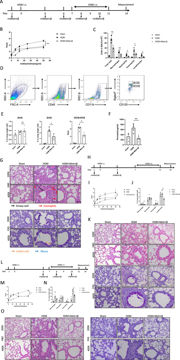
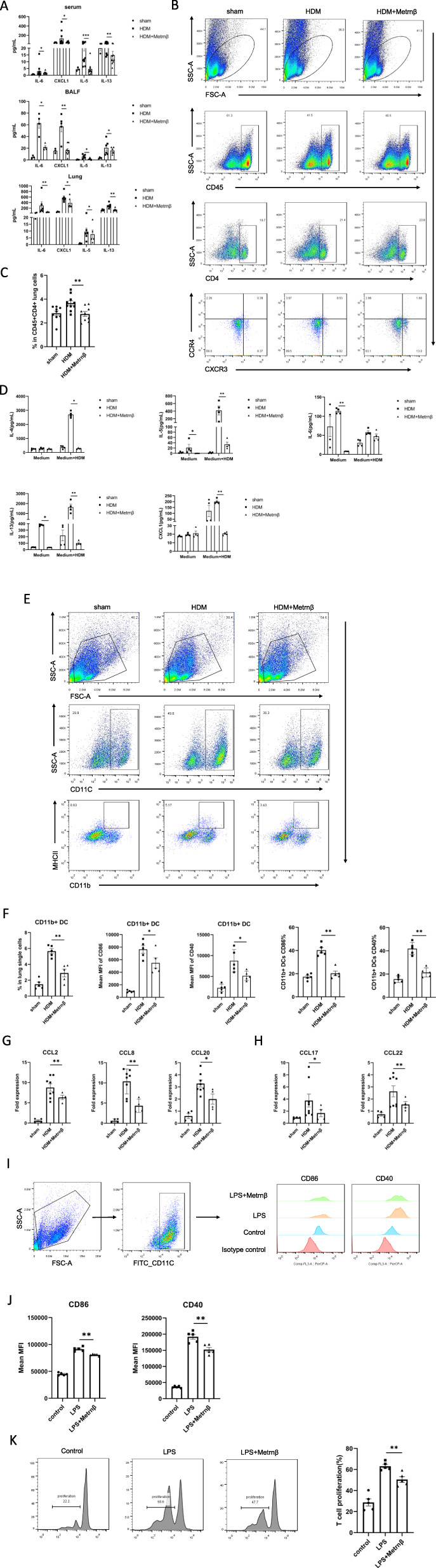
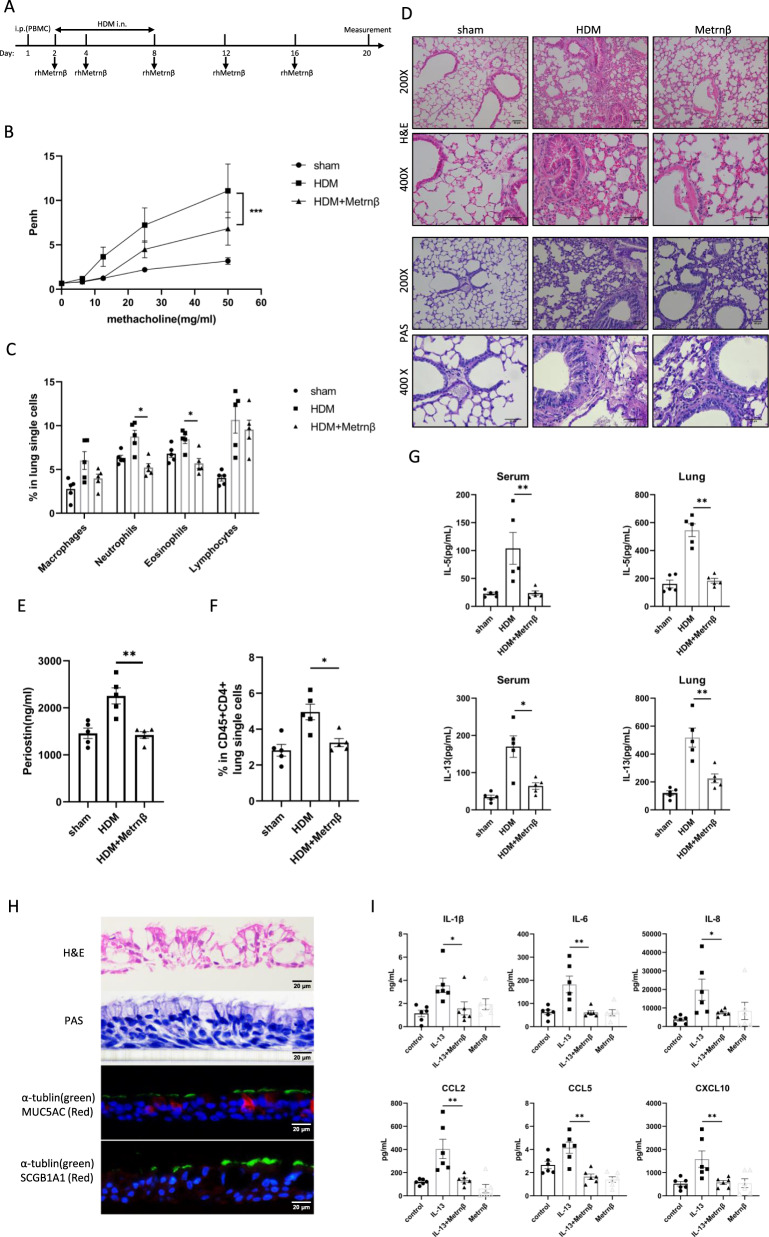
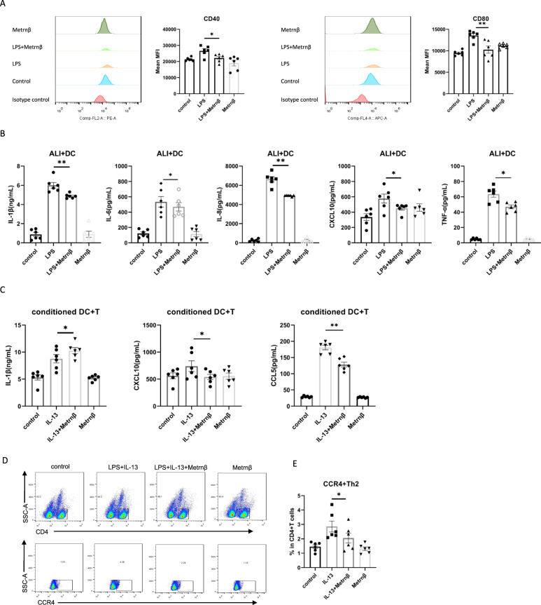
We sought to examine the regulatory effect of Meteorin-β (Metrnβ)/Meteorin like (Metrnl)/IL-41 on lung inflammation in allergic asthma. We found that Metrnβ was elevated significantly in asthmatic patients and in mice with allergic asthma induced by house dust mite (HDM) extract. Upon exposure to HDM, Metrnβ was secreted predominantly by airway epithelial cells and inflammatory cells, including macrophages and eosinophils. The increased Metrnβ effectively blocked the development of airway hyperreactivity (AHR) and decreased inflammatory cell airway infiltration and type 2 cytokine production, which was associated with downregulated DC-mediated adaptive immune responses. Moreover, Metrnβ impaired the maturation and function of bone marrow-derived dendritic cells in vitro. Asthmatic mice adoptively transferred with dendritic cells isolated from Metrnβ-treated allergic mice displayed decreased AHR, airway inflammation, and lung injury. Metrnβ also displayed anti-inflammatory properties in immunodeficient SCID mice with allergic asthma and in in vitro 3D ALI airway models. Moreover, blockade of Metrnβ by anti-Metrnβ antibody treatment promoted the development of allergic asthma. These results revealed the unappreciated protective roles of Metrnβ in alleviating DC-mediated Th2 inflammation in allergic asthma, providing the novel treatment strategy of therapeutic targeting of Metrnβ in allergic asthma.

Keywords: Air–liquid interface; Allergic asthma; Dendritic cells; Eosinophils; Metrnβ.


Conflict of interest statement

The authors declare no competing interests.

Figures

Fig. 1
Fig. 1
Metrnβ expression is increased in allergic asthma. A Circulating Metrnβ concentrations in asthmatic patients (n = 48) and healthy controls (n = 40) were detected by ELISA. BD Metrnβ protein levels in the serum, BAL supernatants and lung tissues of HDM-induced allergic mice assessed with ELISA (n = 5–16). E Representative image of IHC staining for Metrnβ using anti-Metrnβ and isotype IgG in lung sections from asthmatic mice and controls. F IF staining for Metrnβ (green) in lung sections from asthmatic mice and controls. Nuclei were stained with DAPI (blue). G Coimmunofluorescence staining with anti-Metrnβ (red) and anti-e-cadherin (green) in lung sections from asthmatic mice and sham controls and co-IF staining with an anti-Metrnβ (green) antibody and anti-F4/80 (red) or anti-SiglecF (red) antibody to show the colocalization of Metrnβ with macrophages and eosinophils, respectively. H Representative image of flow cytometric analysis of Metrnβ expression on interstitial macrophages and eosinophils in the lungs. I Percentages of Metrnβ-positive macrophages and eosinophils in asthmatic mice detected in (H) (n = 4). Data are representative of three individual experiments. Values are expressed as the mean ± SEM. *P < 0.05; **P < 0.01, and ***P < 0.001 by the Mann–Whitney test
Fig. 2
Fig. 2
Metrnβ controls the severity of allergic inflammation in a mouse model of allergic asthma. A Experimental design for generating an HDM-induced asthma model. rmMetrnβ (1 μg) was injected i.p. 1 day before sensitization and on days 4, 6, 8, and 10. B Airway obstruction of mice treated as in (A) was measured as Penh values to reflect AHR; Penh (% baseline) values were calculated by dividing the methacholine-induced Penh value by the PBS baseline value (n = 4–5). C Total numbers and corresponding cell subtypes were measured in the BALF supernatant of mice treated as in (A) (n = 4–5). D Flow cytometry gating strategies for iEOS and rEOS according to SiglecF and CD125 expression. iEOS were identified as CD45+CD11b+CD125intSiglecFhigh populations, whereas rEOS were defined as CD45+CD11b+CD125intSiglecFint populations. E Detection of iEOS and rEOS in asthmatic lungs with flow cytometry and comparison of the ratio of iEOS/rEOS in asthmatic patients (n = 4–5). F ELISA detection of circulating periostin in asthmatic mice with or without Metrnβ treatment (n = 3–5). G Representative H&E- and PAS-stained lung sections of asthmatic mice treated as in (A). H The experimental timeline for the HDM-induced allergic asthmatic model in which rmMetrnβ (1 μg) was given i.p. 1 day before sensitization and on day 4 of sensitization. I AHR measurement of asthmatic mice treated as in (H) (n = 5). J Total numbers and corresponding cell subtypes were measured in single lung cells from asthmatic mice treated as in (H) (n = 5). K Representative H&E- and PAS-stained lung sections of different groups of mice treated as in (H). L The experimental timeline for the HDM-induced allergic asthma model in which mice were injected i.p. with rmMetrnβ (1 μg) on days 6, 8, and 10. M AHR measurement of asthmatic mice treated as in (L) (n = 5). N Total numbers and corresponding cell subtypes were measured in single lung cells from mice treated as in (L) (n = 5). O Representative H&E- and PAS-stained lung sections of different groups of mice treated as in (L). H&E hematoxylin and eosin, PAS periodic acid-Schiff. Data are representative of at least three individual experiments. Values are expressed as the mean ± SEM. *P < 0.05; **P < 0.01, ***P < 0.001, and ns: not significant
Fig. 3
Fig. 3
Metrnβ diminished the activity of the DC-Th2 axis in allergic asthma. A Cytokine analysis in the serum, BALF and lungs of allergic mice with or without rmMetrnβ (1 μg) treatment (n = 3–10). B Gating strategy and representative flow cytometric plot for the analysis of CD45+CD4+CCR4+CXCR3- Th2 cells in the lungs of asthmatic mice. C Bar chart showing the % of CD45+CD4+CCR4+CXCR3-Th2 cells analyzed in (B) (n = 9–10). D Measurement of cytokines in the supernatants of splenocytes restimulated with HDM extract (30 µg/ml) for 3 days ex vivo. Splenocytes were isolated from mice not given any treatment (sham), HDM-induced allergic mice (HDM), and rmMetrnβ-treated allergic mice (HDM + Metrnβ) (n = 4). E Gating strategy and representative flow cytometric dot plots for the analysis of CD11b+ DCs in the lungs of allergic mice. F Bar chart showing the numbers of CD11b+ DCs in (E), the mean MFIs of CD86 and CD40 on CD11b+ DCs, and the percentages of CD86+ and CD40+ cells in total CD11b+ DCs (n = 5). G, H mRNA expression of chemokines in the lungs of allergic mice measured using qRT-PCR (n = 4–8). I BMDCs isolated from wild-type BALB/C mice were cultured at a density of 2 × 105/ml and stimulated with LPS (2 µg/ml) for 48 h with or without rmMetrnβ (100 ng/ml) treatment. Cells were collected for the detection of DC maturation markers including CD86 and CD40 (n = 5). The gating strategy and representative graphs for the detection of DC maturation markers are displayed. J Calculation of the MFIs of CD86 and CD40 in (I) is summarized (n = 5). K CD11c+ BMDCs (2 × 104) from wild-type BALB/C mice were pulsed with LPS (2 µg/ml) and incubated at a 1:5 ratio with CFSE-labeled splenic CD4+ T cells (1 × 105) for 4 days with or without rmMetrnβ (100 ng/ml) treatment. Cell proliferation is presented as the percentage of proliferated CD4+ T cells determined using flow cytometric analysis (n = 5). Data are representative of at least three individual experiments. Values are expressed as the mean ± SEM. *P < 0.05 and **P < 0.01
Fig. 4
Fig. 4
In vivo effects of CD11c+ DCs isolated from rmMetrnβ-treated mice on the induction of type 2 airway inflammation. A Experimental protocol and timepoint for adoptive transfer in our study. CD11c+ BMDCs were isolated from HDM-sensitized and challenged mice with or without rmMetrnβ treatment and enriched by magnetic cell sorting. CD11c+ BMDCs (3 × 105) were adoptively transferred into recipient mice via i.n. administration 2 h before HDM challenge on day 7, followed by sequential challenge with HDM from day 7 to day 11. Mice were sacrificed for analysis on day 14. B AHR measurement before sacrifice using whole-body plethysmography (n = 5). C Analysis of lung inflammatory cells, including macrophages, neutrophils, eosinophils, and lymphocytes, by flow cytometry, as represented by a bar chart (n = 5). D Representative H&E- and PAS-stained lung sections showing lung damage, airway thickness, and mucus production in allergic SCID mice. E Bar chart showing the percentage of CD45+CD4+CCR4+CXCR3- Th2 cells in the lungs using flow cytometric analysis (n = 5). F Measurement of Th2-related cytokines, including IL-5 and IL-13, in the serum and lungs by using a Milliplex assay (n = 5). Data are representative of two individual experiments. Values are expressed as the mean ± SEM. *P < 0.05 and **P < 0.01
Fig. 5
Fig. 5
Metrnβ reversed the hyperresponsiveness of humanized HDM-induced allergic mice and the 3D ALI model. A Experimental protocol and timepoints for the humanized allergic asthmatic mouse model. SCID mice were reconstituted by i.p. transplantation of purified human PBMCs (2 × 107) obtained from asthmatic or control subjects on day 1, followed by i.n. treatment with 50 μg HDM on days 2, 4, and 8 with or without rhMetrnβ (1 μg) injected i.p. on days 2, 4, 8, 12, and 16. All mice were sacrificed for analysis on day 20. B AHR measurements of mice before sacrifice using whole-body plethysmography (n = 5). C Counts of inflammatory cells in samples of single lung cells determined with flow cytometry, as represented by a bar chart (n = 5). D Representative H&E- and PAS-stained lung sections of humanized HDM-induced allergic mice. E ELISA detection of the serum periostin concentration (n = 5). F Flow cytometric analysis of CD45+CD4+CCR4+CXCR3- Th2 cells in the lungs (n = 5). G Measurement of Th2-related cytokines, including IL-5 and IL-13, in the serum and lungs by ELISA (n = 5). H Representative images showing the differentiation of primary HBEpiCs into ALI cultures with H&E staining and mucus-producing goblet cells identified by PAS staining, as well as IF staining for ciliated cells (α-tubulin, green), goblet cells (muc5AC, red), and club cells (SCGB1A1, red). Nuclei were stained with DAPI (blue). I Differentiated ALI cultures (28 days) were treated with rhIL-13 (100 ng/ml) for 24 h, with or without rhMetrnβ pretreatment for 2 h, and differential expression of cytokines and chemokines in the basal compartment of the ALI system was quantified by using a CBA (n = 6). Data are representative of two individual experiments. Values are expressed as the mean ± SEM. *P < 0.05, **P < 0.01, and ***P < 0.001
Fig. 6
Fig. 6
Effects of Metrnβ on epithelium–dendritic cell crosstalk in allergic asthma. ALI models derived from HBEpiCs were cultured in PneumaCult™-ALI medium on the basal side for 28 days to obtain a differentiated cell population with a mucociliary phenotype. HMDCs (5 × 105) were cocultured with an ALI model with or without rhMetrnβ (100 ng/ml) pretreatment for 2 h, followed by stimulation with LPS (2 µg/ml) or LPS (2 µg/ml) + IL-13 (100 ng/ml) for 48 h. A DCs were collected for flow cytometric analysis of the surface expression of CD40 and CD80 (n = 6). The relative expression of CD40 and CD80 is shown as the mean fluorescence intensity (MFI). B The contents of the basal compartment were collected for evaluation of IL-1β, IL-6, IL-8, CXCL10, and TNF-α by using a CBA (n = 6). CE Conditioned DCs from the control, IL-13, IL-13+Metrnβ, and Metrnβ-stimulated ALI+DC coculture groups were separately cocultured with human CD4+ T cells for 48 h. After centrifugation, the supernatant was analyzed for IL-1β and CXCL10 release by using a CBA. CCL5 in the supernatant was quantitated by using ELISA (C), and cells were collected for T cell polarization analysis by using flow cytometry. A representative gating image and distinct expression of CD4+CCR4+ T cells in different groups are shown in (D) and (E) (n = 6). Data are representative of two independent experiments. Values are expressed as the mean ± SEM. *P < 0.05 and **P < 0.01
Fig. 7
Fig. 7
Metrnβ blockade aggravated AHR and allergic inflammation in asthma. A Experimental protocol and timepoints for the treatment of mice with an anti-Metrnβ monoclonal antibody. B AHR measurement before mouse sacrifice using whole-body plethysmography (n = 5). C Counting of inflammatory cells in single lung cells via flow cytometry, as represented by a bar chart (n = 4). DE Flow cytometric analysis of CD11b+ DCs and CD45+CD4+CCR4+CXCR3- Th2 cells in the lungs (n = 4). F Representative H&E- and PAS-stained lung sections of allergic mice treated with the anti-Metrnβ antibody or an isotype control. GH Measurement of cytokines and chemokines, including IL-6, CXCL1, IL-5, IL-13, IL-33, and CCL11, in the serum and lungs by a Milliplex assay (n = 4–5). Data are representative of two independent experiments. Values are expressed as the mean ± SEM. *P < 0.05 and **P < 0.01

References

    1. Schatz M, Rosenwasser L. The allergic asthma phenotype. J Allergy Clin Immunol Pract. 2014;2:645–8. - PubMed
    1. Christiansen SC, Zuraw BL. Treatment of hypertension in patients with asthma. N Engl J Med. 2019;381:1046–57. - PubMed
    1. Hekking PW, Wener RR, Amelink M, Zwinderman AH, Bouvy ML, Bel EH. The prevalence of severe refractory asthma. J Allergy Clin Immunol. 2015;135:896–902. - PubMed
    1. Zheng T, Yu J, Oh MH, Zhu Z. The atopic march: progression from atopic dermatitis to allergic rhinitis and asthma. Allergy Asthma Immunol Res. 2011;3:67–73. - PMC - PubMed
    1. Mishra A, Yao X, Saxena A, Gordon EM, Kaler M, Cuento RA, et al. Low-density lipoprotein receptor-related protein 1 attenuates house dust mite-induced eosinophilic airway inflammation by suppressing dendritic cell-mediated adaptive immune responses. J Allergy Clin Immunol. 2018;142:1066–79.e1066. - PMC - PubMed

Publication types