. 2022 May 24;145(4):1464-1472.
doi: 10.1093/brain/awab388.

Molecular trans-dural efflux to skull bone marrow in humans with CSF disorders

Affiliations

Molecular trans-dural efflux to skull bone marrow in humans with CSF disorders

Geir Ringstad et al. Brain. .

Abstract

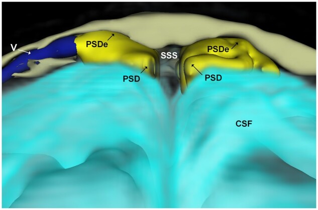
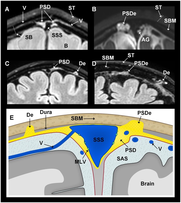
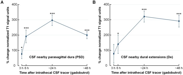
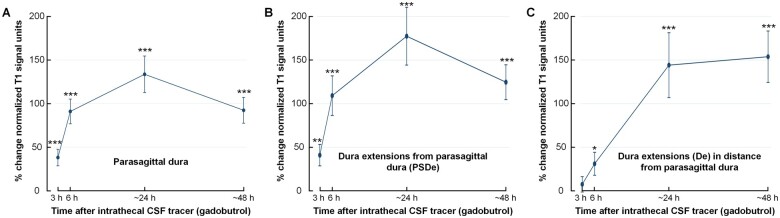
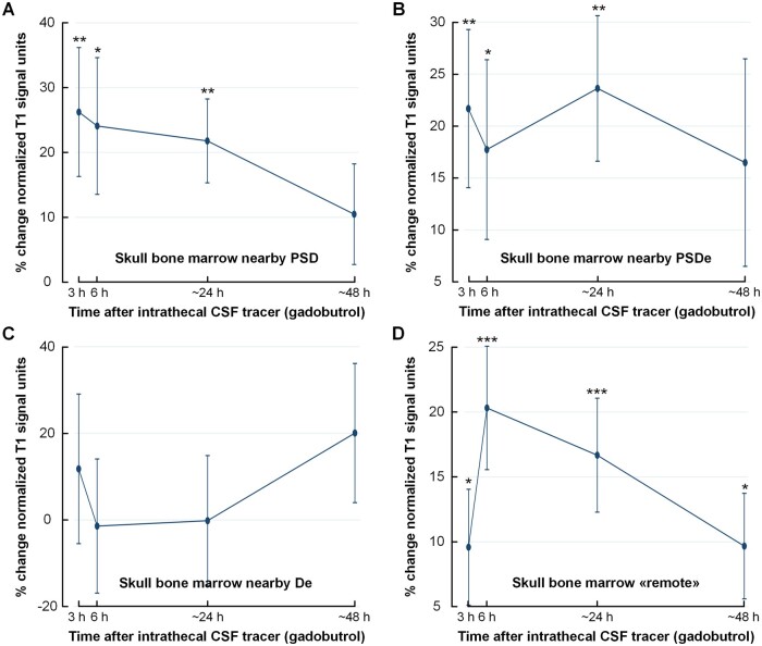
Dural sinuses were recently identified as a hub for peripheral immune surveillance of brain-derived antigens cleared through CSF. However, animal studies have also indicated that substances and cells may enter the intracranial compartment directly from bone marrow. We used MRI and a CSF tracer to investigate in vivo whether intracranial molecules can move via dura to skull bone marrow in patients with suspicion of CSF disorders. Tracer enrichment in CSF, dural regions and within skull bone marrow was assessed up to 48 h after intrathecal administration of gadobutrol (0.5 ml, 1 mmol/ml) in 53 patients. In participants diagnosed with disease, tracer enrichment within diploe of skull bone marrow was demonstrated nearby the parasagittal dura, nearby extensions of parasagittal dura into diploe, and in diploe of skull bone remote from the dura extensions. This crossing of meningeal and skull barriers suggests that bone marrow may contribute in brain immune surveillance also in humans.

Keywords: bone marrow; dural lymphatic vessels; immune system; parasagittal dura.


Figures

Figure 1
Figure 1
3D representation (coronal plane) illustrating dural extensions from PSD into the intradiploic bone marrow space at skull vertex. The PSD (yellow) directly overlies the CSF within the subarachnoid space (turquoise). An intradiploic vein (V; dark blue) traverses within the skull bone marrow (beige) adjacent to the PSD. It can be noted how the PSD extends into the intradiploic bone marrow space (PSDe). The different colours of CSF and PSD/PSDe reflect different tracer enrichments. Image: Tomas Sakinis, MD.
Figure 2
Figure 2
Patients with dural extensions into bone marrow of skull, either directly or in distance from PSD (all coronal sections at skull vertex). In this study, we examined three main intradural locations, namely the parasagittal dura (PSD), parasagittal dura extensions (PSDe) into dipolic area of skull bone marrow and dural extensions (De) into skull bone marrow, but not in direct continuity with PSD. Examples are retrieved from four different patients. (A) The PSD is a loose soft tissue matrix adjacent to the superior sagittal sinus (SSS), here visualized by T2-FLAIR. (B) The PSDe cross the inner table into the diploe of skull, and are separated from the subarachnoid space (SAS). It is different from macroscopically visible arachnoid granulations (AG) bulging into the sinus. Here, both PSDe and AG are enriched by CSF tracer (T1-BB, 24 h). (C) In distance from PSD, De into diploic area of skull bone marrow (SBM) may be seen (T2-FLAIR). (D) The size and shape of parasagittal and dural extensions (PSDe, De) vary considerably (T2-FLAIR). (E) Cartoon illustrating the examined anatomical structures beyond the superficial tissue (T2-FLAIR). MLV = meningeal lymphatic vessels; ST = superficial tissue. Illustration in E: Øystein Horgmo, University of Oslo.
Figure 3
Figure 3
Tracer enrichment over time in CSF of subarachnoid space nearby the regions of interest. Tracer enrichment in CSF of subarachnoid space (A) nearby PSD (n = 47) and (B) and nearby De to diploe of skull bone in distance from PSD (n = 12). The percentage change in normalized T1-BB signal, indicative of tracer enrichment, was highly significant in both locations (linear mixed model analysis; *P < 0.05, **P < 0.01, ***P < 0.001), with peak at 24 h. Trend plots presented by mean and error bars as standard errors.
Figure 4
Figure 4
Tracer enrichment in different intradural locations. Tracer (gadobutrol) enrichment over time within (A) PSD (n = 47), (B) PSDe (n = 27) and (C) within De into diploe of skull bone marrow in distance from PSD (n = 12). The plots show tracer enrichment as a percentage change in normalized T1-BB signal units. The signal change, indicative of tracer enrichment, was highly significant for all locations (linear mixed model analysis; *P < 0.05, **P < 0.01, ***P < 0.001), with peak at 24 h. Trend plots presented by mean and error bars as standard errors.
Figure 5
Figure 5
Tracer enrichment within skull bone within different regions of interest. Tracer (gadobutrol) enrichment over time in different regions of skull bone marrow. (A) Skull bone marrow nearby PSD (n = 47). (B) Skull bone marrow nearby dural extensions from parasagittal dura (PSDe) to diploe (n = 27). (C) Skull bone marrow nearby dural extensions (De) to diploe in distance from the PSD (n = 12). (D) Skull bone marrow in distance from visible intradural tracer enrichment, denoted ‘remote’ (n = 47). The plots show the percentage change in normalized T1-BB signal units. The signal change, indicative of tracer enrichment, was highly significant for several locations (linear mixed model analysis; *P < 0.05, **P < 0.01, ***P < 0.001), with peak at 24 h. Trend plots presented by mean and error bars as standard errors.

Similar articles

Cited by

References

    1. Abbott NJ. Evidence for bulk flow of brain interstitial fluid: Significance for physiology and pathology. Neurochem Int. 2004;45(4):545–552. - PubMed
    1. Louveau A, Smirnov I, Keyes TJ, et al. . Structural and functional features of central nervous system lymphatic vessels. Nature. 2015;523(7560):337–341. - PMC - PubMed
    1. Aspelund A, Antila S, Proulx ST, et al. . A dural lymphatic vascular system that drains brain interstitial fluid and macromolecules. J Exp Med. 2015;212(7):991–999. - PMC - PubMed
    1. Jacob L, Boisserand LSB, Geraldo LHM, et al. . Anatomy and function of the vertebral column lymphatic network in mice. Nat Commun. 2019;10(1):4594. - PMC - PubMed
    1. Ringstad G, Eide PK.. Cerebrospinal fluid tracer efflux to parasagittal dura in humans. Nat Commun. 2020;11(1):354. - PMC - PubMed

Publication types